Palaemon or Macrobrachium malcolmsonii
(The Indian Freshwater Prawn)
- Palaemon (The Freshwater Prawn) is studied in the Indian Universities as a typical representative of the class Crustacea.
- Common Indian species 🡪 Palaemon carcinus ; Palaemon malcolmsonii
Systematic Position of Palaemon
Phylum : Arthropoda
Class : Crustacea
Subclass : Malacostraca
Order : Decapoda
Suborder : Macrura
Family : Palaemonidae
Genus : Palaemon
Habit and Habitat of Palaemon
- Palaemon inhabits freshwater streams, rivers, ponds and lakes.
- It is a nocturnal creature, hiding at the bottom during the day and coming to the surface at night in search of food.
- It is omnivorous, feeding on small organisms, like algae, mosses, minute insects, debris, e.t.c.
- In a desperate attempt to escape from the enemy’s grasp, it can shed off one or more of its appendages. This phenomenon is known as autotomy.
- During the breeding period (May – July) the female is seen carrying a large number of eggs between its abdominal appendages.
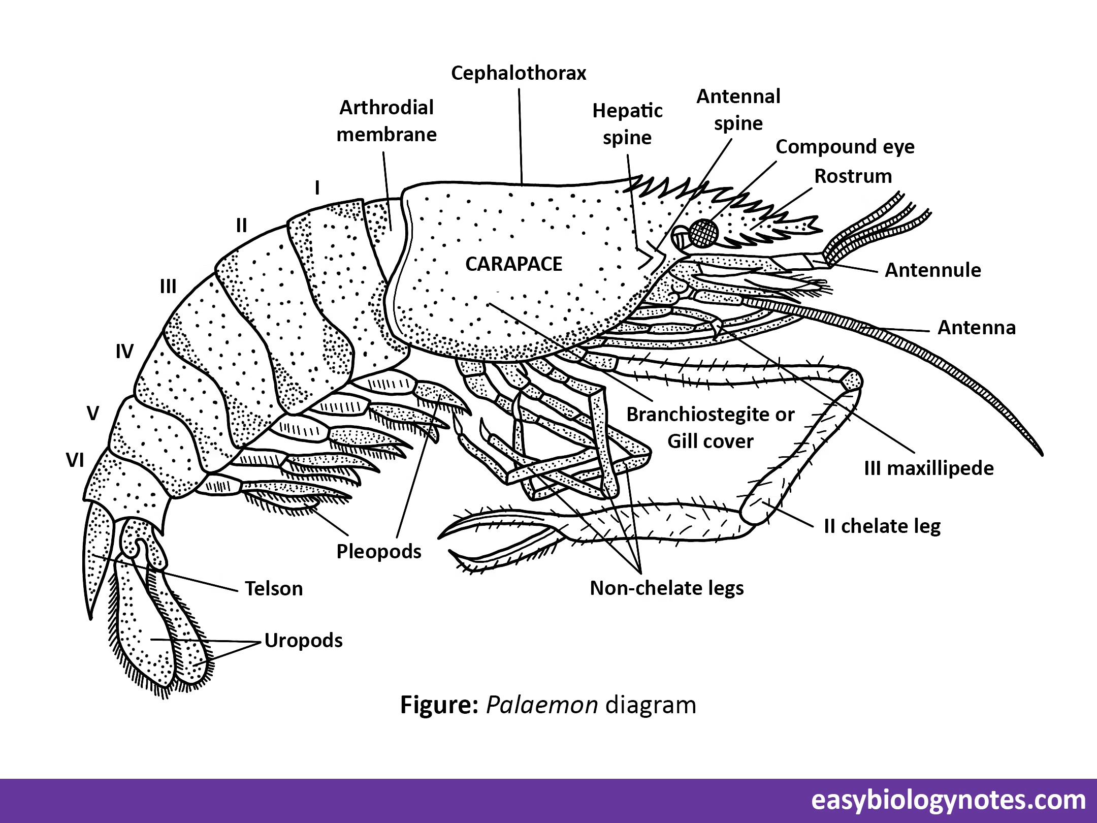
External Morphology of Palaemon
Shape and Size
- Body is elongated, more or less spindle-shaped and bilaterally symmetrical. It offers less resistance in swimming.
- Size of an adult varies from species to species.
Palaemon malcolmsonii, now Macrobrachium malcolmsonii, found in Central India and Tamil Nadu, measures 25 to 40 cm in length. - The giant prawn Palaemon carcinus from Kerala is upto 90 cm long. While the dwarf prawn Palaemon lamarrei, found almost throughout India, is 2,5 to 5 cm long.
Colouration
- Young stages are transparent and white, but the adults are differently tinted according to the species.
- Usual colour is dull pale-blue or greenish with brown orange-red patches.
- Preserved specimens are deep orange.
Segmentation and Body Divisions
- Body of an adult prawn is distinctly divided into 19 segments or somites, all bearing jointed appendages.
- The segments are arranged into two main regions: an anterior cephalothorax (fused head-thorax) and a posterior abdomen.
1. Cephalothorax
- Cephalothorax is large, rigid, unjointed and more or less cylindrical in shape.
- It consists of 13 segments.
- Cephalothorax is formed by the union of two regions: Head and Thorax.
- Head consists of 5 segments while thorax includes 8 segments, all bearing jointed appendages.
2. Abdomen
- Well developed abdomen is jointed, unlike cephalothorax.
- It is composed of 6 distinct movable segments, and a terminal conical piece, the tail plate or telson, which is not considered a segment because of post-segmented origin.
- Abdominal segments are dorsally rounded, laterally compressed and normally bent under the cephalothorax, so that the animal looks like a comma (,) in shape.
- The abdomen looks almost circular in a cross section. Each abdominal segment carries a pair of jointed appendages, called pleopods or swimmeret.
External apertures
- The slit-like mouth opens mid-ventrally at the anterior end of cephalothorax.
- Anus is a longitudinal aperture lying ventrally at the base of telson.
- Paired renal apertures open on raised papillae on inner surface of coxae of antennae.
- Paired female genital apertures in female open on the inner surface of coxae of the third pair of walking legs.
- Paired male genital apertures in the male are situated on the inner surface of coxae of the fifth pair of walking legs.
- There are two minute openings of statocysts, one lying in a deep depression dorsally on the basal segment (precoxa) of each antennule.
Exoskeleton
- Body and appendages are covered by a hard protective calcareous shell or exoskeleton.
It is composed of chitinous cuticle which becomes variously tinted by the deposition of lime salts and sclerotin. - The exoskeleton comprises several hardened plates, called sclerites.
Appendages
- Each segment bears a pair of jointed appendages. Thus, there are 19 pairs of appendages in Palaemon.
- They show considerable variations depending on the functions they perform. However, they all are of a biramous (bi, two + ramus, branch) type (with the exception of antennules which are uniramous), as they are built on the same fundamental embryonic origin.
- In prawn, there are 19 pairs of appendages, 13 pairs of cephalothorax and 6 in abdomen. Cephalothoracic appendages further include 5 pairs of anterior cephalic appendages and 8 pairs of posterior thoracic appendages
Cephalic Appendages
- There are 5 pairs of cephalic or head appendages.
- Beginning from the anterior end of head they are the antennules, antennae, mandibles, maxillulae, and maxillae. Antennules and antennae are pre-oral, while mandibles, maxillae and maxillulae are post-oral.
(a) Antennules – The antennules are attached, one on either side, below the bases of eye-stalks. The feelers of antennules bear sensory setae and are tactile in function.
(b) Antennae – The antennae lie, one on either side, just below the antennules. The antennae are sensory, excretory and balancing in function.
(c) Mandibles – The two mandibles are strong calcified bodies, lying one on either side of the mouth. Mandibles constitute the biting jaws and are masticatory in function.
(d) Maxillulae – These are small, thin and leaf-like appendages. Maxillulae help in the manipulation of food.
(e) Maxillae – These are also thin and leaf-like mouth appendages. Maxillae help in respiration and in the manipulation of food.
Thoracic Appendages
- There are 8 pairs of thoracic appendages. These are differentiated into anterior 3 pairs of maxillipedes (Gr., maxilla – jaw + podos – food) or foot-jaws and posterior 5 pairs of paraeopods or walking legs.
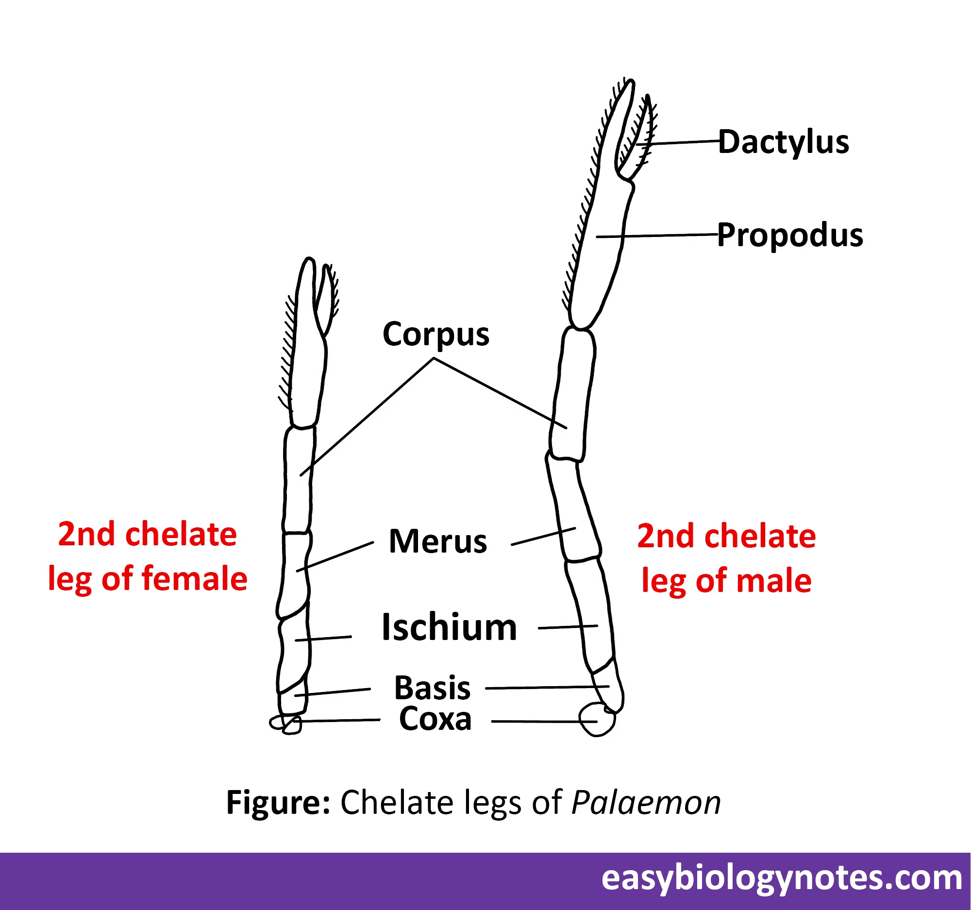
Walking legs – In the first and second pairs of walking legs, propodus is prolonged beyond its articulation with dactylus, so that the two podomers work against the other like blades of a pair of forceps and from a chela or pincer. Such legs are termed as chelipeds or chelate legs.
- They are used to grasp food and pass it on to the mouth. They also serve as organs of offence and defence.
- The second chelate legs in male are larger and more powerful than in female.
- The third, fourth and fifth pairs of legs are non-chelate and typical.
- In female, each third leg bears a female reproductive aperture on the inner side of the coxa.
While in male, each fifth leg bears a male genital aperture on the anthrodial membrane between the leg and thorax.
Abdominal Appendages
- Abdomen bears 6 pairs of abdominal appendages, one pair in each of its segments.
- First 5 pairs are the swimming pleopods or swimmerets, used as paddles, while the 6th pair are the uropods which, along with the post-segmental telson, form the tail fin. All these appendages are of simple biramous type.
Reproductive System of Palaemon
Sexual Dimorphism
The sexes are separate (dioecious) and sexual dimorphism is well marked.
- Male is bigger in size than female.
- The male possesses a narrower abdomen than female.
- In male, second chelate legs are longer, stronger and more spiny than in female.
- In male, paired genital openings lie on the coxae of 5th pair of walking legs, while they lie on the coxae of 3rd pair of legs in female.
* A pair of gonads are similar in position, shape, size and general deposition in both the sexes.
[A] Male Reproductive System
1) Testes – The two testes are soft, white and elongated bodies which fuse at their anterior ends to form a common lobe.
- They enclose between them a gap for the passage of the cardio-pyloric strand connecting heart to pyloric stomach.
- Histologically, each testis consists of a large number of coiled, narrow and thin walled seminiferous tubules embedded in connective tissue.
- The cavity of each tubule is lined by a single layer of germinal epithelium, the cells of which undergo spermatogenesis to form spermatozoa.
- A mature sperm consists of a rounded cytoplasmic body, containing a large, dark, crescentic nucleus, and a tail-like blunt process.
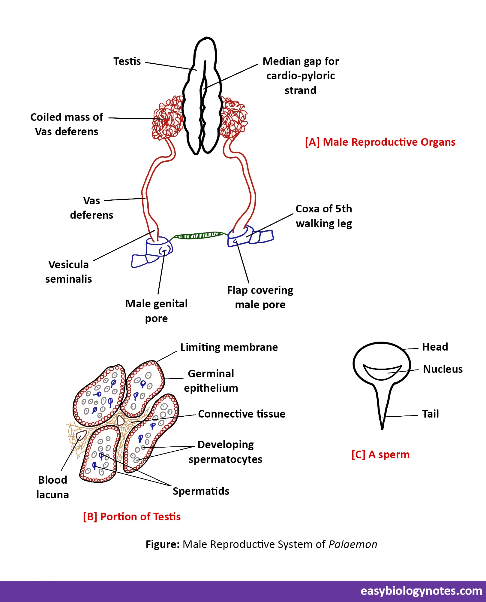
2) Vas deferentia – A long, coiled and narrow tube, the vas deferens, arises from each testis near its posterior end.
3) Vesicula seminalis – Each vas deferens reaching ventrally near the base of fifth walking leg, swells to form a club-shaped vesicula seminalis.
- These store spermatozoa in the form of white bodies called spermatophores.
- Each vesicula seminalis or seminal vesicle opens to the exterior through a male genital aperture situated on the inner side of coxa of fifth walking leg of its side.
- Each male genital aperture is covered by a small flap of integument.
[B] Female Reproductive System
1) Ovaries – The two ovaries are white, compact and sickle-shaped bodies touching each other at both the ends but leaving a gap in the middle for the passage of the cardio-pyloric strand. Each ovary is enclosed within a membranous capsule.
2) Oviducts – A short, wide and thin walled tube, the oviduct, originates from the outer middle border of each ovary. It runs vertically downwards to open through a female genital aperture on the inner side of the coxa of third walking leg of its side.
