What is Excretion and Excretory System?
The process of removal of chemical waste (mainly nitrogenous) from the body is known as excretion and the organ system involved is called Excretory System.
- Excretion plays an important role in maintaining the homeostatic condition of the body.
Excretory Products
- CO2 and Water
- Nitrogenous Waste Products
- Nitrogen containing waste products are produced by the breakdown of excess amino acids or proteins or nucleic acids (DNA and RNA) .
- The nitrogenous waste poroducts are of the following three types –
- Ammonia
- It is produced by oxidative deamination of amino acids.
- It is highly toxic.
- The animals which secrete ammonia are called ammonotelic.
- The excretion of ammonia is called ammonotelism.
- Example – Hydra , Bony fishes, tailed amphibians .
- Urea
- It is produced in the liver by the deamination of excessive amino acids (ammonia and CO2) .
- It is poisonous and if allowed to accumulate in the blood to a certain level may cause death.
- It is highly soluble in water and is removed from the body in the form of urine.
- The animals which excrete urea are termed as ureotelic.
- The removal of urea is called ureotelism.
- Example – Vertebrates and Invertebrates.
- Uric acid
- It is least toxic.
- Removal of uric acid is called uricotelism.
- The animals which excrete uric acid are called uricotelic.
- It is chief excretory product in insects, reptiles, birds and desert animals.
- Ammonia
- Mineral Salts
Human Excretory System
- All the body organs which help the animal in excretion collectively constitute the excretory system.
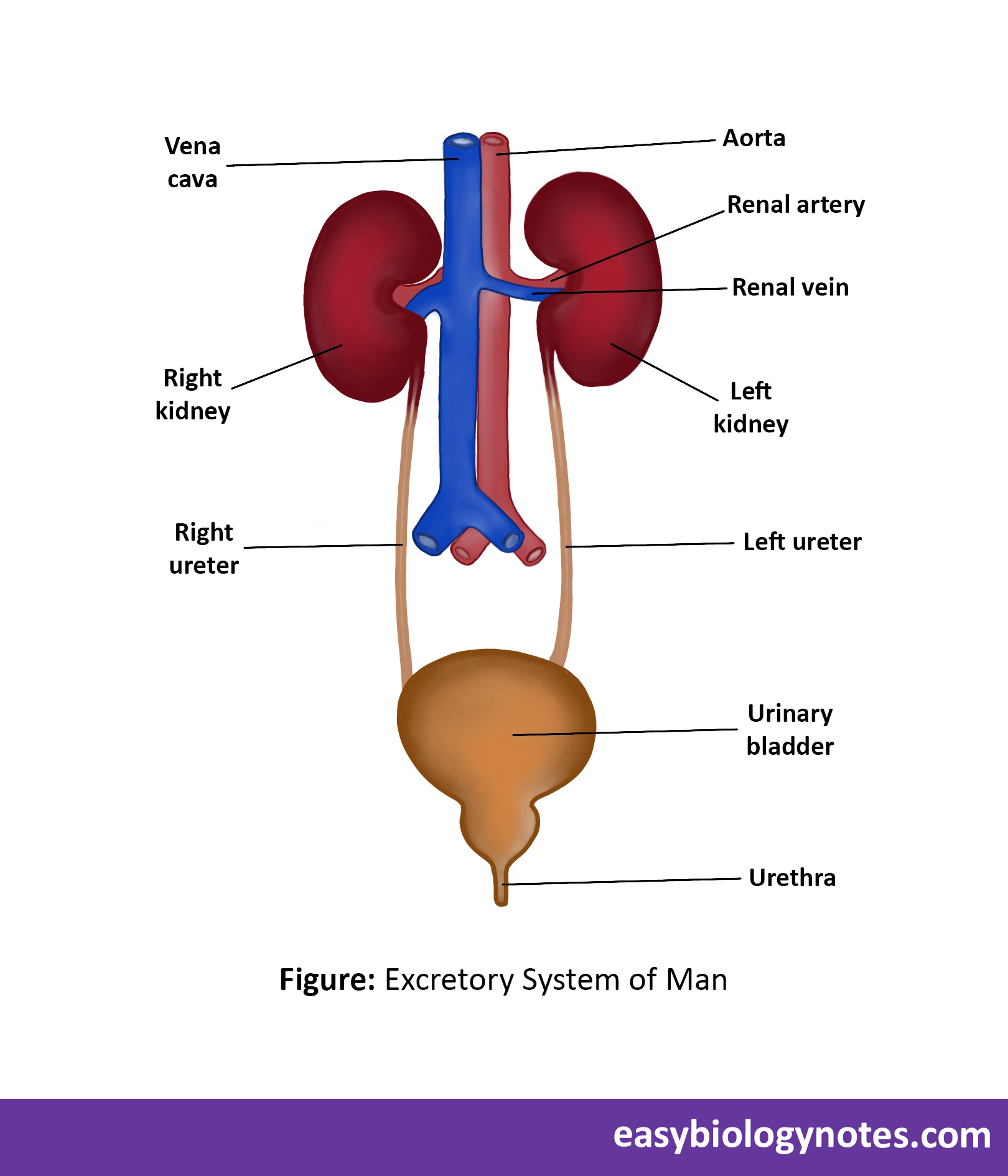
- Excretory system consists of following organs –
- A pair of kidneys
- A pair of ureters
- Urinary Bladder
- Urethra
Lets discuss each of the above mentioned organs of the Excretory System in detail-:

-
Kidneys
- Most important organ of the excretory system.
- Shape and size
- Each kidney is reddish-brown bean shaped slightly flattened organ.
- Each kidney is about 10cm long, 6cm wide and 3.5 cm thick.
- It has a bean shaped structure.
- Location
- They are present just below the stomach , on the dorsal side, one on either side of the vertebral column protected by the last two ribs.
- Right side of kidney is at slightly lower level than the left one because the right kidney is pushed downwards by the large right liver lobe.
- External structure of kidney
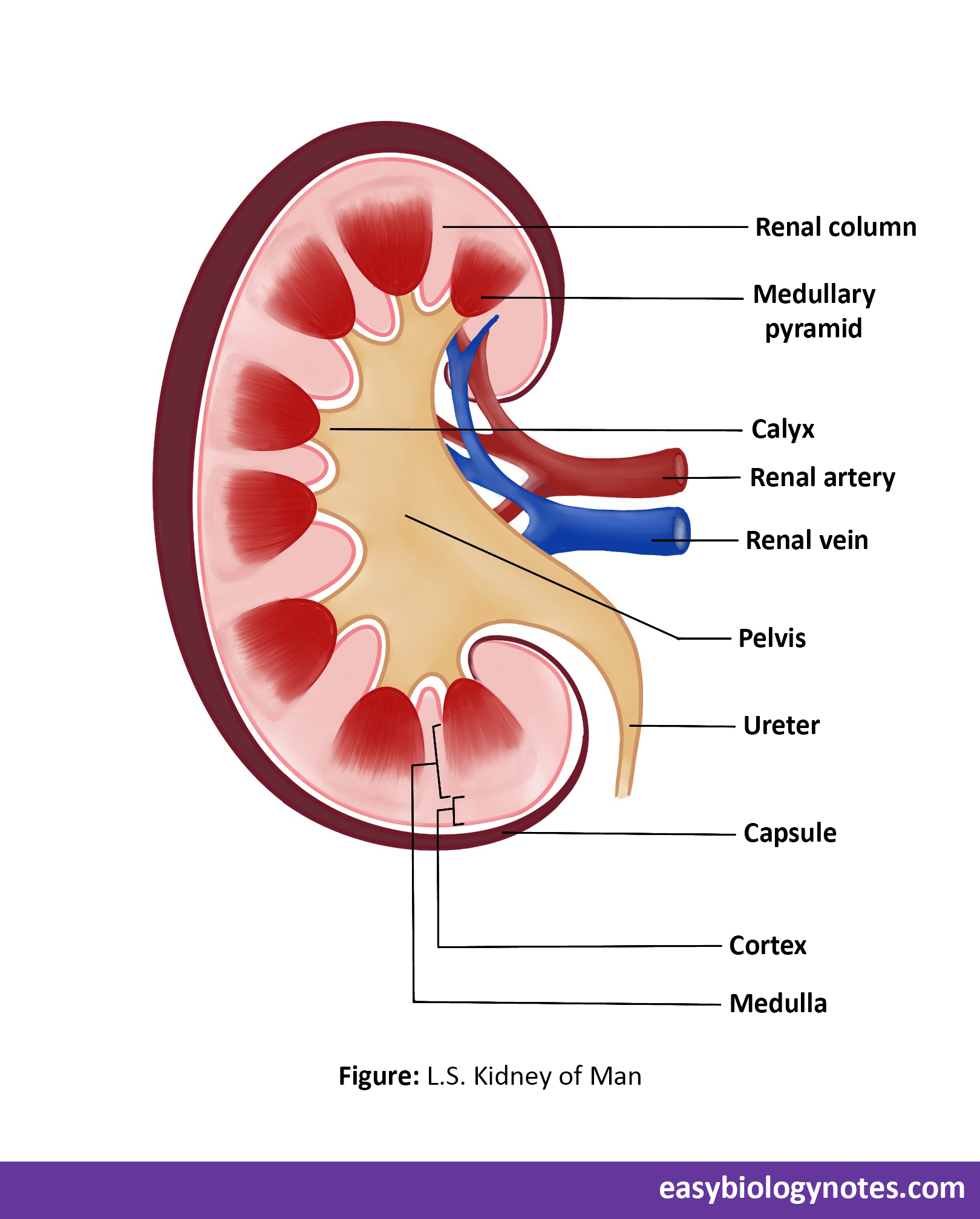
- Outer margin of kidney is convex and inner margin is concave.
- In the middle of inner concavity is present a notch called hilus or hilum.
- The renal artery , renal vein , nerve fibre , lymph vessels and uterus enter or leave the kidney through hilus.
- Internal structure of kidney
- Externally, kidney is bounded by a thin sheet of white fibrous tissue called capsules (renal capsule).
- Each kidney contain about 1.25 million tiny structural and functional units of kidney which are highly coiled tubules called nephrons or uriniferous tubules.
- Kidney is differentiated into two regions – the outer darker region is called cortex and the inner light region is called the medulla.

Structure of nephrons or uriniferous tubules
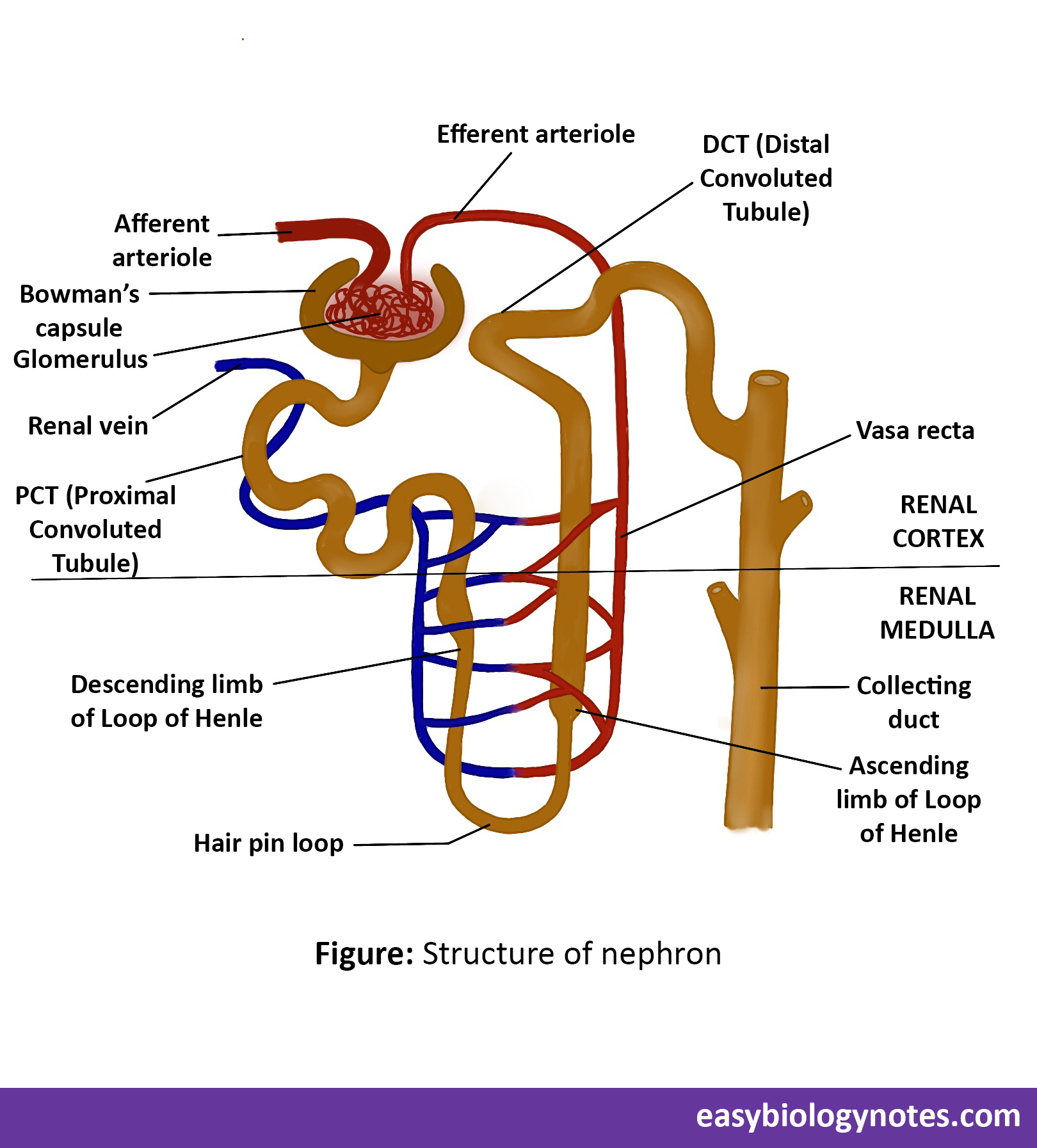
- Each nephron is highly coiled tube like structure and is differentiated into two parts –:
- Malpighian capsule or renal corpuscle – It is differentiated into two parts and lies into cortical region of kidney .
a. Bowman’s capsule – It is a thin double – walled single cell thick epithelium, cup-like structure.
b. Glomerulus – It is a knot of capillaries present in the cup of Bowman’s capsule.- It is formed by the capillaries of incoming blood vessel (afferent arteriole) and that of outgoing blood vessel (efferent arteriole).
- Malpighian capsule or renal corpuscle – It is differentiated into two parts and lies into cortical region of kidney .
-
- Renal tubules
The Bowman’s capsule leads into a coiled part of nephron called Renal tubule which is further differentiated into three parts :
- PCT (Proximal Convoluted Tubule) – Lies in the cortex region.
- Loop of Henle – It is middle U- shaped part of Renal Tubule and is shaped like a hair pin.
- It consists of descending limb, hair pin loop and ascending limb.
- It is found in medulla of kidney.
- Distal Convoluted Tubule (DCT)
Collecting duct or collecting tubule – The open ends of so many nephrons open into a wider tube called collecting tubule or collecting duct. The collecting duct receives the contents of many kidney tubules and pores it as urine in the pelvis of the kidney.

Note -:
Each single tubule = 4 to 5 cm long
Total number of tubules in both kidneys is around 2 million.
Total length of all tubules together = < 60 km. This great length provides a huge surface area for reabsorption.
Blood supply to the kidney tubules
- A large branch of abdominal aorta called renal artery supplies blood to each kidney.
- Each renal artery branches and rebranches several times to give rise to arterioles.
- Afferent arteriole enters Bowman’s capsule and breaks into a number of capillaries which forms a knot like mass of capillaries called glomerulus.
- The reuniting capillaries of glomerulus form the efferent arteriole which after emerging from Bowman’s capsule runs a short distance and breaks up into a secondary capillary network called vasa recta.
- The capillaries of this system again unite to form renal venule which unites again and again with other veins and ultimately forms the renal vein.
2. Ureter
-
- Ureters are a pair of long narrow and whitish tubes.
- Each ureter is about 10-12 inches in length and less than half a inch in diameter.
- In kidney, it expands forming a funnel shaped basin called the renal pelvis.
- Ureter carry urine from kidney to urinary bladder.
3. Urinary bladder
-
- It is pear-shaped, highly distensible, sac-like reservoir present in the pelvic region.
- The opening of bladder into urethra is guarded by a sphincter muscle.
- It can hold about 0.5 to 1 liter of urine.
4. Urethra
-
- Urethra is a narrow tube which extends from the floor of the bladder to the exterior.
Mechanism of Urine Formation or Physiology of Excretion
Physiology of Excretion can be studied into two steps :
-
Chemical Steps (Formation of Urine)
- In the liver, the oxidative deamination of excess amino acids releases ammonia.
- Ammonia due to its strong basic nature is highly toxic therefore quickly changed into urea by combining with CO2.
-
Mechanical steps
- The Urea produced in the liver cells is transported to kidneys by the way of blood for elimination. In the kidneys, the urea, along with small uric acid, mineral salts, excess of water, pigments, e.t.c. is converted into a faint yellow coloured urine.
- The formation of Urine is completed by three processes :-
-
Ultrafiltration
- The blood flows through the glomerulus under great pressure and the reason for this greater pressure is that the efferent arteriole is narrower than the afferent arteriole thus a pressure is build up in the glomerulus capillaries.
- This high pressure (hydrostatic pressure) causes the liquid part of the blood to filter out from the glomerulus under tremendous pressure which is known as ultrafiltration.
-
Or ,
Ultrafiltration is the process of filtration of the liquid part of blood out from the Glomerulus into the Bowman’s capsule because of hish pressure set up in the glomerulus due to the differences in the diameter of the afferent and efferent arteriole.
-
-
-
- During ultrafiltration almost all liquid part of the blood, that is plasma, along with urea, uric acid , glucose, amino acid, water, inorganic salts and other substances like pigments comes out of the glomerulus and passes into the funnel shaped cavity of Bowman’s capsule. This filtered out liquid is called glomerular filtrate or nephric filtrate or ultrafiltrate.
- The blood that passes into the efferent arteriole is left with only blood proteins, corpuscles and fats that cannot pass through the walls of capillaries and hence the blood is relatively thick.
- Urine produced from glomerulur filtrate after reabsorption per day is equal to 1 – 1.5 litre.
2. Selective reabsorption
- The process by which only useful substances are reabsorbes from the nephric filtrate into the blood running through secondary capillary network is called selective reabsorption.
- PCT reabsorbs entire glucose, most of the water, some inorganic salts like sodium, potassium and chloride ions.
- Loop oh Henle reabsorbs water and sodium ions.
- DCT and collecting tubules reabsorbs some amount of water and remaining chlorides.
3. Tubular Secretion
- The secretion of harmful substances from the blood into the nephric filtrate through the walls of DCT is called tubular secretion.
- Some substances such as creatinine, potassium ions, ammonia, and drugs are added to nephric filtrate by the cells of distal convoluted part.
- The filtrate left after reabsorption and tubular secretion is called urine.
- Selective reabsorption and tubular secretion help to maintain proper acid base balance of the body.
-
-
Properties and composition of normal urine
| Physical Properties of Normal Urine | Chemical properties of normal Urine | |
|
Compound
Besides these substances, the urine may also contain certain hormones, certain medicines like antibodies and excess vitamins |
g/litre2.30.7
1.5 2.6 9.0 2.5 1.8 0.6 0.2 1.7 1.5 |
|
||
|
||
|
||
|
||
Urine excretion
- Final urine passes into collecting ducts, to the renal pelvis, and through the ureter to the urinary bladder by ureteral peristalsis (waves of constriction into uterus) and due to gravity.
- The process of passing out urine is called micturition.
- Egestion – The removal of undigested food and other debris from the intestine is called egestion.
Regulation of Urine Output
- The posterior lobe of pituitary gland secretes a hormone called antidiuretic (ADH) which controls the concentration of urine by water reabsorption.
- If ADH secretion is reduced there is an increased production of urine. This is called diuresis.
- Substance that increases the formation of urine are called diuretics. Example – Tea, Coffee, Alcohol, Beer, etc.
Osmoregulation
- Process of maintaining the water acid salt contents constant in the body is called osmoregulation.
- Kidneys in addition to excretory functions also acts as osmoregulatory organs.
- Thus, kidney while removing waste like urea from the blood also regulates its composition, i.e. , the percentage of water and salts.
- Osmoregulation is extremely important , eg. , if the osmotic pressure of the tissues and blood is different water diffuses from low osmotic pressure to high osmotic pressure. This helps in the normal working of the body.
- Drinking enough water directly or through food helps the kidney in their proper working. The frequency of urination is fewer times in summer than in winter. The reason is that in summer there is a considerable loss of water through precipitation and the kidneys have to reabsorb more water from the urine making it more concentrated whereas in winter there is no sweating and most of the excess water is eliminated fron the body in the form of urine.
- In cholera, patient suffers from vomiting and diarrhoea because of the inability of intestine to absorb water into the blood. To maintain water contents constant, kidneys absorb almost all the water from the urine even with some urea. Ultimately the patient may die due due to poisoning by the accumulation of high quantities of urea in the body (uremia).
Disorders of Kidneys
- Hematuria (Blood in urine) – In this disease blood is passed out along with urine. The usual cause of this disease is the toxin produced in certain types of fever and infection in the urinary tract, kidney stone, or tumour.
- Uremia – In this disease excessive urea is retained in the blood because of the failure of nephrons to extract urea from the blood.
- Glycosuria (Glucose in the urine) – In this disease the level of blood sugar rises and excess glucose passes with urine due to diabetes mellitus (Sugar diabetes).
- Kidney stones
- Kidney stones are formed by the precipitation of uric acid and salts like calcium oxalate. These may be forms in the kidneys, pelvis, ureters, urinary tubules and urethra.
- They are removed surgically but some types of kidney stones may also be dissolved.
- Nephritis – Inflammation of the kidneys is called nephritis.
Dialysis – The removal of unwanted substances from the body through artificial kidney is called dialysis.
Functions of kidneys in Excretory System
Kidneys perform many functions to maintain the internal environment constant (homeostasis). These functions are -:
- Excretion of nitrogenous waste products – Nitrogenous waste products like urea, uric acid are eliminated from the body in the form of urine by kidneys
- Regulation of water balance – If the water is excess in the body large amounts of dilute urine are produced whereas during deficiency of water hypertonic (solution which has higher solute composition , here urine) urine is produce.
- Regulation of acid – base balance.
- Elimination of substances like pigment, drugs, e.t.c.
- Regulation of blood pressure – Kidneys regulate the arterial blood pressure by secreting a hormone called rennin.
- Kidneys maintain fluid homeostasis in the body – By elimination of nitrogenous waste products, excess of water, excess of salts and other unwanted substances from the blood, the kidneys maintain the volume composition, pH and osmotic pressure of the blood.