- All the parts of the body that help in the transportation of various materials in the body, collectively constitute the Circulatory System.
- In higher and multicellular organisms there is a well organized transport system of the body by wich the blood is being circulated within a closed system being circulated within a closed system under different pressure gradient created by the pumping mechanisms where the heart acts as the central pump.
- The main components of the circulatory system are -:
- A fluid transport medium (blood and lymph)
- Controlling centre – The Heart
- Path through which transport medium circulates- The blood vessels and the lymph vessels.
The Human Heart
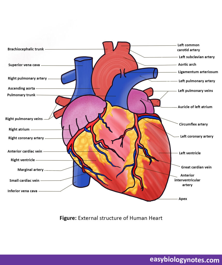
- Human Heart is a reddish brown pumping centre.
- It pumps and collects the blood to and from the various body parts.
- SIZE – About the size of a person’s close fist and weighs about 300 grams.
- SHAPE – It is a broad conical structure.
- Upper side of the heart is broad called base and lower pointed side is called the apex.
- POSITION – Present right in the centre between the two lungs in the thoracic cavity above the diaphragm.
- Well protected by the bones that forms thoracic cage.
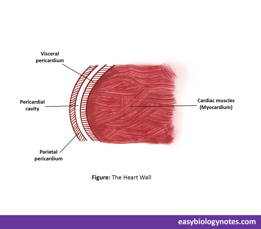
Pericardium and wall-layers of the heart
- The heart is covered by a two layer sac known as pericardium.
- Inner layer is called visceral layer of pericardium and the outer layer is called parietal layer of pericardium.
- In between these two layers , a narrow space known as pericardial cavity is present which is full of a self secreted fluid called pericardial fluid.
- The pericardial fluid performs the following functions-:
- Protects the heart from any kind of injury or shock.
- It keeps the tissue of heart moist for proper functioning.
- It acts as lubricant and reduces friction for the beating of heart.
Structure of Human Heart
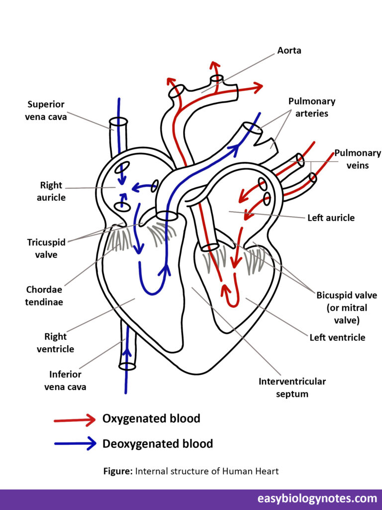
- The human heart consists of four chambers which are as follows -:
- Two atria or auricles
- Two ventricles
- Atria or Auricles
- Auricles are a pair of thin walled chambers present towards the base of the heart separated from each other by inter-auricular septum thus prevents mixing of oxygenated and deoxygenated blood.
- They are thin walled because they have to push the blood into the ventricles present just below them.
- Ventricles
- Ventricles are a pair of very thich walled chambers present towards the apex of the heart, which remains separated from each other by inter ventricular septum.
- Left ventricles has the thickest walls as it has to push the blood to the distinct parts of the body such as those of the feet.
- From the right upper corner of the left ventricle starts a large tubular aorta that supplies blood to all body parts.
- From the left upper corner of right ventricle starts a large tubular pulmonary arch which supplies deoxygenated blood to the lungs.
Blood vessels entering and leaving the heart, their apertures and valves
- Radius blood vessels that enter or leave the heart are called great blood vessels.
- Apertures are the openings through which blood vessels enter or leave the heart.
- Apertures are guarded by flap like structures known as valves. They allow unidirectional flow of blood through the heart.
Blood vessels entering the heart
- In the right auricle
a) Superior (Anterior) Vena Cava: It brings deoxygenated blood from upper parts of the body – head , neck, chest and arms. It is not guarded by any valve. It is also called precaval.
b) Inferior (Posterior) Vena Cava: It brings deoxygenated blood from lower parts of the body – abdomen and legs which opens into the right auricle through an aperture which is guarded by Valve of Eustachius.
* Right auricle opens into the right ventricle through right auriculo-ventricular aperture which is guarded by three leaf- like flaps(cusps) known as tricuspid valve. - In the left auricle
Oxygenated blood from the lungs is collected by pulmonary veins (4 in number, 2 from each lung).
*Left auricle opens into the left ventricle through auriculo-ventricular aperture which is guarde by two leaf-like flaps(cusps) called bicuspid valve.
Blood vessels leaving the heart
- Pulmonary arch / trunk
It arises from the right ventricle and carries deoxygenated blood to the lungs for oxygenation. Opening of pulmonary arch is guarded by a valve called pulmonary semi-lunar valve. - Aorta
It arises from the left ventricle and supplies oxygenated blood to all the body parts.Its opening is guarded by a valve called aortic semi-lunar valve - Coronary arteries
The vessels that deliver blood to the muscles (Myocardium) of the heart itself are called coronary arteries.
Blockage of any coronary artery or its branches causes deadening of correspomding area of heart muscles. It is called myocardial infarction or heart attack.
It causes pain in chest and is called angina pectoris.
Heartbeat
- The rhythmic contraction and relaxation of heart chambers (i.e. , auricles and ventricles) by the contraction and relaxation of cardiac muscles is known as the heartbeat.
- Heartbeat consists of two phases-:
- The contraction phase (which is called systole)
- The relaxation phase (which is called diastole)
Circulation of blood through the heart
- Deoxygenated blood from all the body parts is collected into the right auricle by superior and inferior vena cava.
- Simultaneously oxygenated blood from the lungs is collected into the left auricle by the way of four pulmonary veins.
- When the auricles are full of blood, a wave of contraction (systole) passes into them. The ventricles at this time are relaxing and are empty. Therefore the blood from the auricles passes into the ventricles easily. The two cuspid opens and allow the blood to flow into the ventricles.
- Now the wave of contraction passes into the ventricles and the contract. At this time the flaps of the two cuspid valves get tightened and closes the passage, thus preventing the backflow of blood to the auricles.
- Blood from the right ventricle passes into the pulmonary arteries whereas from the left ventricle into the aorta by opening of the semilunar valves.
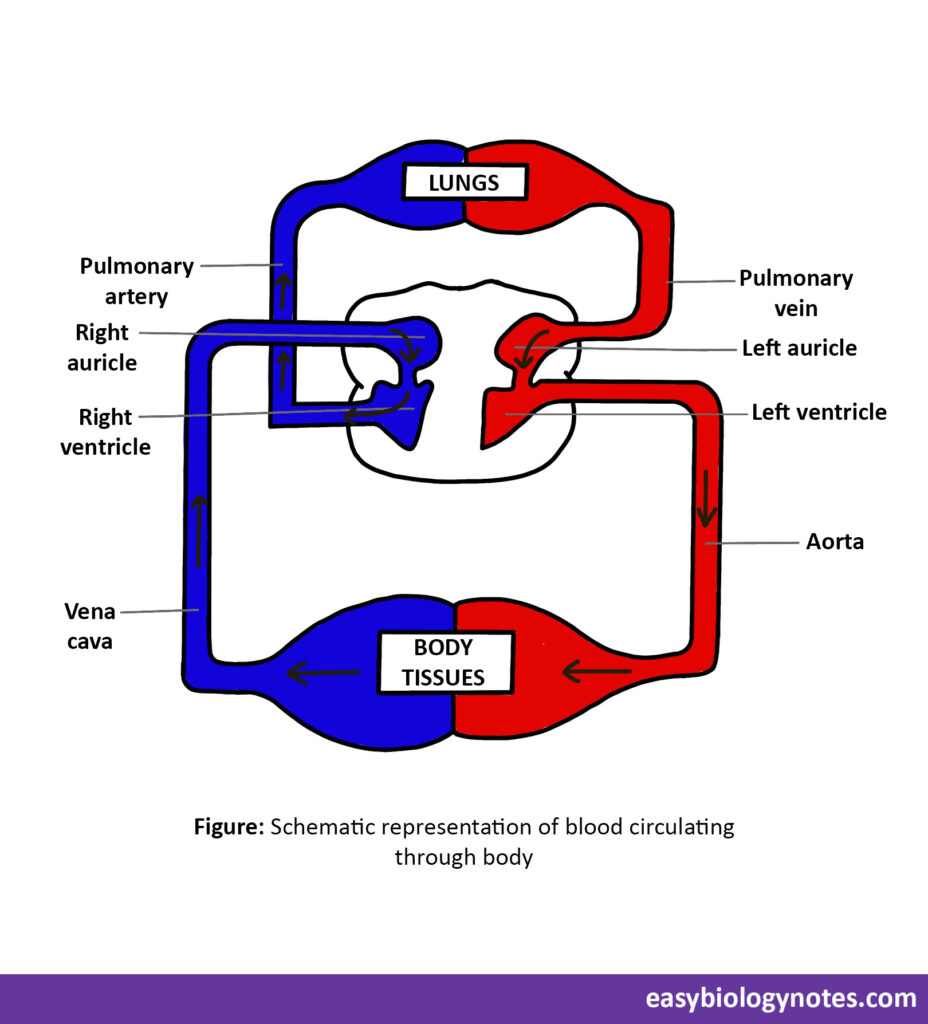
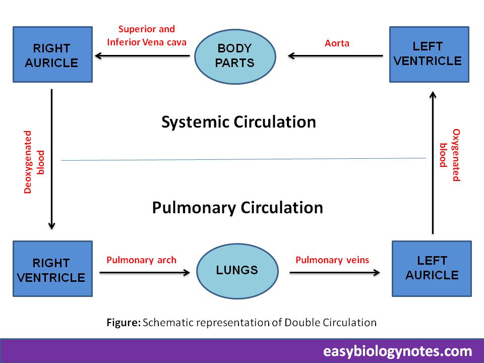
Double Circulation
Double Circulation- The Blood has to pass atwice through the heart so as to circulate once through the body. This type of circulation is called double circulation.
It is as follows-:
- Pulmonary circulation– Circulation of deoxygenated blood from the right ventricle to the left auricle through lungs is called pulmonary circulation.
- Systemic circulation – The circulation of oxygenated blood from the left ventricle to the right auricle through various body parts or systems is called systemic circulation.
Cardiac Output or Heart Output
- The amount of blood pumped by the heart per minute is called cardiac output or heart output.
- Amount of blood pumped in 1 beat = 70ml
- Number of beats per minute = 72
- Therefore, amount of blood pumped in 1 minute= 70*72 = 5040ml = 5 litre (approx)
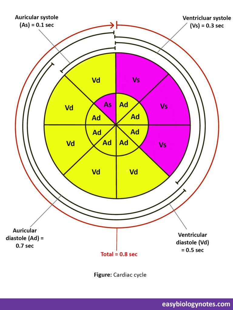
Cardiac Cycle
- The Cardiac events that occur from the beginning of one heartbeat to the beginning of the next is called cardiac cycle.
- Human heart beats 72 times in one minute so one heart beat is completed in 0.8 seconds.
- The cardiac cycle or 1 heart beat consists of following three phases :
- Auricular systole – During this phase auricles contract and push blood into corresponding ventricles. It is completed in 0.1 seconds.
- Ventricular systole – During this phase ventricles contract and push the blood into pulmonary arteries and aorta. It is completed in 0.3 seconds.
- Joint diastole – During this phase all the four chambers of the heart are relaxed. It extends for 0.4 seconds.
Origin , conduction and regulation of heart beat
Nodal tissue – Heart Muscles consist of specialized muscle fibres which have the properties of neurons and are responsible for initiation and transmission of cardiac impulses known as nodal tissue.
They comprise of the following structures :
- Sino – atrial node (S-A node)
Present in the wall of right auricle near the opening of superior vena cava. It is also called as pacemaker as heartbeat first originates at the S-A node and maintains the basic rhytm of the heartbeat. - Auricular – ventricular node (A-V node)
Arises from S-A node and present in the right side of inter auricular septum. - A-V bundle or Bundle of His
It arises from A-V node and passes downwards in the inter ventricular septum and then divides into left and right bundle branches. - Purkinje fibres
Arises from Bundle of His and forms a network which spreads throughout the walls of ventricles.
Conduction of Heartbeat
- The heartbeat starts at S-A node causes the contraction of auricles and spreads in all directions.3
- It stimulates the A-V node which also sends wave of contraction to the ventricles through the Bundle of His and Purkinje fibres.
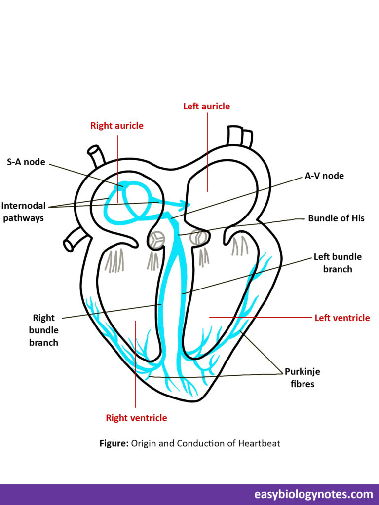
*Heartblock – If the Bundle of His does not function properly the ventricles will not contract. This condition is called heartblock.
Frequently Asked Questions (FAQs)
Question – Name the two heart sounds and how they are produced.
Answer – Lubb and dup are the two heart sounds .
- The first sound i.e., lubb is a long booming sound which is produced by the closure of auriculo – ventricular valves soon after ventricular systole.
- The second sound i.e., dup , is a short sharp sound which is produced due to the closure of semi lunar valves.
Question – What is the technique used to register heart sounds?
Answer – PHONOCARDIOGRAPHY
Question – Instrument used to measure heart beat is ___________
Answer – STETHOSCOPE