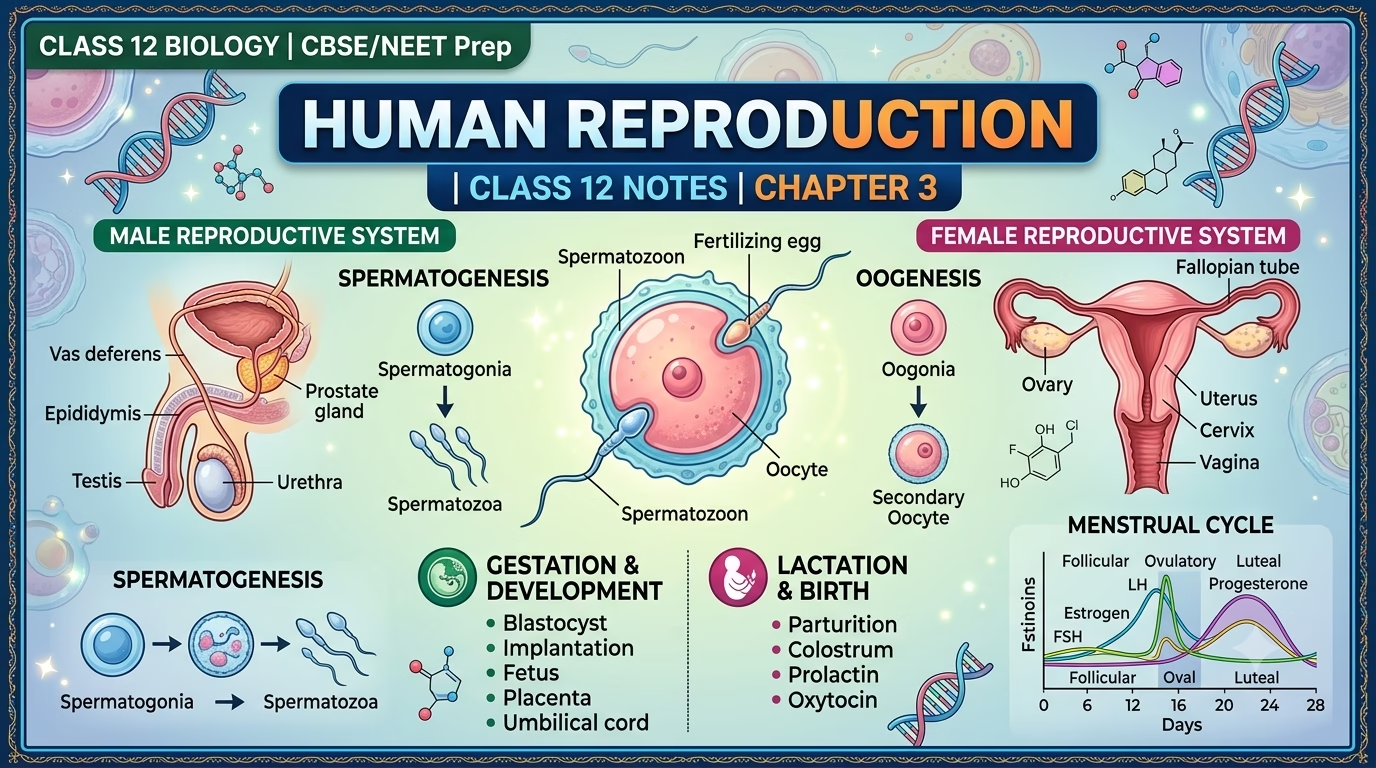
Human Reproduction Class 12 Notes Biology- A complete, exam-ready explanation of the male and female reproductive systems, gametogenesis, the menstrual cycle, fertilisation, pregnancy, and parturition — with diagrams, key terms, and quick-revision points for Class 12 Biology students.
Introduction to Human Reproduction
Human beings reproduce sexually and are viviparous, meaning offspring develop inside the mother’s body and are born alive rather than hatching from eggs. This topic is one of the most important chapters in Class 12 Biology (NCERT) and forms the foundation for later chapters on reproductive health and genetics.
The reproductive phase in humans begins at puberty and involves a coordinated sequence of biological events:
- Gametogenesis – formation of sperm and ova
- Insemination – deposition of semen into the female reproductive tract
- Fertilisation – fusion of sperm and ovum
- Implantation – attachment of the embryo to the uterine wall
- Gestation – development of the foetus inside the uterus
- Parturition – the process of childbirth
Let’s look at each stage in detail, starting with the reproductive organs themselves.
The Male Reproductive System
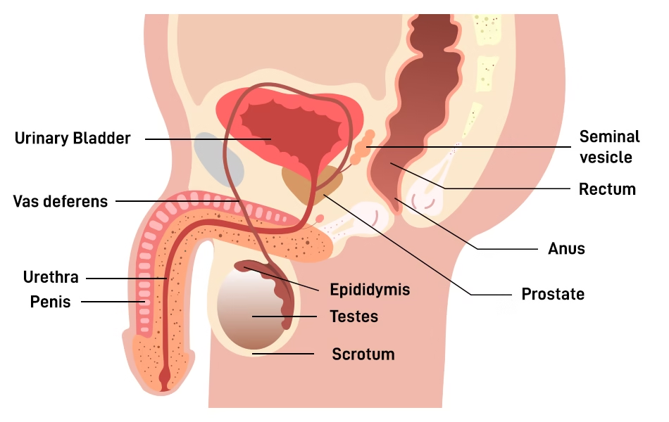
The male reproductive system is located in the pelvic region and consists of a pair of testes, a network of accessory ducts and glands, and external genitalia. Together, these structures produce, mature, and transport sperm.
Testes
- The testes lie inside a skin-covered pouch called the scrotum, which lies outside the abdominal cavity. This external position keeps the testes about 2–2.5°C cooler than body temperature, which is essential for normal sperm production.
- Each testis measures roughly 4–5 cm in length and 2–3 cm in width, and is divided internally into about 250 compartments called testicular lobules.
- Each lobule contains 1–3 highly coiled seminiferous tubules, the actual sites of sperm formation.
- The seminiferous tubules are lined by two cell types:
- Male germ cells (spermatogonia) – undergo meiosis to eventually form sperm.
- Sertoli cells – often called “nurse cells,” they nourish the developing germ cells and secrete factors that support spermatogenesis.
- The region outside the seminiferous tubules is called the interstitial space. It contains Leydig cells (interstitial cells), which synthesise and secrete androgens (male sex hormones, mainly testosterone).
Accessory Ducts
Sperm produced in the seminiferous tubules travel through a series of ducts before leaving the body:
Rete testis → Vasa efferentia → Epididymis → Vas deferens → Ejaculatory duct → Urethra
- The rete testis collects sperm from the seminiferous tubules and channels them into the vasa efferentia.
- The vasa efferentia lead into the epididymis, a long coiled tube where sperm mature and are stored.
- The epididymis continues as the vas deferens, which loops over the urinary bladder.
- The vas deferens joins the duct of the seminal vesicle to form the short ejaculatory duct, which opens into the urethra.
- The urethra originates at the urinary bladder, passes through the penis, and opens to the exterior via the urethral meatus.
Accessory Glands
- A pair of seminal vesicles
- Prostate gland
- A pair of bulbourethral glands (Cowper’s glands)
The secretions of these three glands combine with sperm to form seminal plasma. This fluid nourishes sperm, provides a medium for their motility, and helps neutralise the acidity of the vagina during intercourse.
The Female Reproductive System
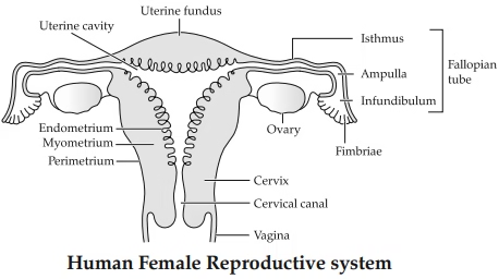
The female reproductive system also lies in the pelvic region and includes a pair of ovaries, a pair of oviducts (fallopian tubes), the uterus, cervix, vagina, external genitalia, and the mammary glands (which, while not strictly part of the reproductive tract, play a vital role in nurturing offspring).
Ovaries
- The ovaries are the primary female sex organs. They produce the ovum (egg) and secrete ovarian hormones — chiefly oestrogen and progesterone.
- Each ovary is 2–4 cm long and is attached to the pelvic wall and to the uterus by ligaments.
- The ovary is covered by a layer of epithelium and contains the ovarian stroma, divided into:
- Peripheral cortex – contains ovarian follicles at various stages of development.
- Inner medulla – contains blood vessels, nerves, and connective tissue.
Oviducts (Fallopian Tubes)
- Each oviduct is about 10–12 cm long and extends from the ovary to the uterus.
- The end closer to the ovary is funnel-shaped and called the infundibulum, which has finger-like projections called fimbriae that help sweep the released ovum into the tube.
- The infundibulum leads into the wider ampulla — the usual site of fertilisation — and then narrows into the isthmus, which joins the uterus.
Uterus
- Also known as the womb, the uterus is pear-shaped and is supported by ligaments attached to the pelvic wall.
- The uterine wall has three layers:
- Perimetrium – the outer covering
- Myometrium – the thick middle layer of smooth muscle, responsible for the strong contractions of labour
- Endometrium – the innermost lining, which undergoes cyclical changes during the menstrual cycle and is where the embryo implants
Cervix and Vagina
- The cervix is the lower, narrow part of the uterus that opens into the vagina.
- Together, the cervix and vagina form the birth canal.
External Genitalia (Vulva)
- Mons pubis – a cushion of fatty tissue covered by skin and pubic hair, overlying the pubic bone
- Labia majora – thick folds of skin extending from the mons pubis, surrounding the vaginal opening
- Labia minora – thinner folds of skin lying beneath the labia majora
- Hymen – a thin membrane that partially covers the vaginal opening
- Clitoris – a small, highly sensitive erectile structure at the junction of the labia minora
Mammary Glands
- Present in all female mammals; paired, glandular structures.
- Each breast contains 15–20 mammary lobes, each made up of clusters of alveoli that secrete milk.
- Milk from the alveoli drains into mammary tubules, which unite to form a mammary duct.
- Several mammary ducts join to form a mammary ampulla, a small storage reservoir connected to the lactiferous duct, which opens at the nipple.
Gametogenesis: Formation of Sperm and Ova
Gametogenesis is the process by which the testis and ovary produce gametes:
- Spermatogenesis in males (production of sperm)
- Oogenesis in females (production of ova)
Spermatogenesis
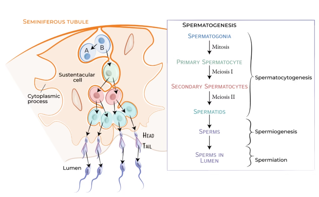
- Spermatogonia (immature diploid germ cells) line the inner walls of the seminiferous tubules and multiply by mitosis.
- Some spermatogonia, called primary spermatocytes, periodically undergo meiosis I, producing two haploid secondary spermatocytes.
- The secondary spermatocytes undergo meiosis II, producing four haploid spermatids.
- Spermatids are transformed into functional, motile sperm through a process called spermiogenesis.
- After spermiogenesis, the sperm heads become embedded in the Sertoli cells and are eventually released into the lumen of the seminiferous tubule by a process called spermiation.
Hormonal control of spermatogenesis:
- Spermatogenesis begins at puberty due to a significant rise in Gonadotropin-Releasing Hormone (GnRH) from the hypothalamus.
- GnRH stimulates the anterior pituitary to release two gonadotropins: Luteinising Hormone (LH) and Follicle-Stimulating Hormone (FSH).
- LH acts on Leydig cells, stimulating them to secrete androgens, which in turn stimulate spermatogenesis.
- FSH acts on Sertoli cells, stimulating factors that assist spermiogenesis.
Structure of a Sperm
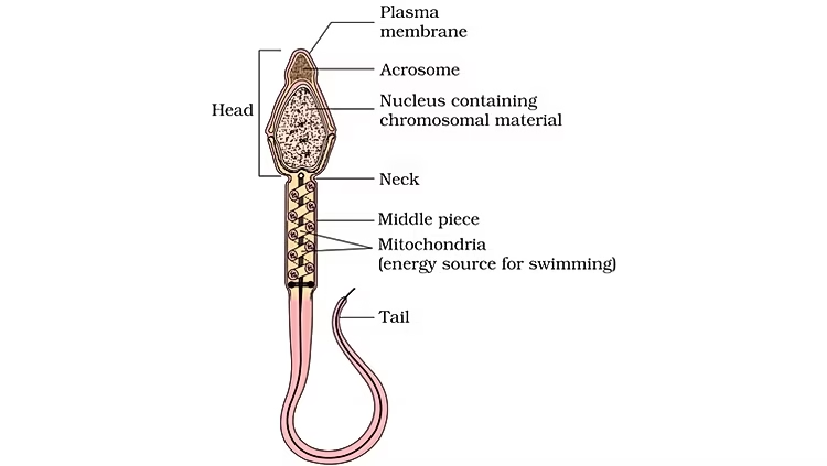
A mature human sperm has four main regions, all enclosed within a single plasma membrane:
| Region | Key Features |
| Head | Contains a haploid nucleus and a cap-like acrosome filled with enzymes essential for penetrating the ovum during fertilisation |
| Neck | Short connecting region containing centrioles |
| Middle piece | Packed with mitochondria, which generate the energy (ATP) needed for sperm motility |
| Tail | A flagellum that provides the propulsive, whip-like movement of the sperm |
Once released from the seminiferous tubules, sperm are transported through the accessory ducts. Secretions from the epididymis, vas deferens, seminal vesicles, and prostate gland are essential for the final maturation and motility of sperm.
Oogenesis
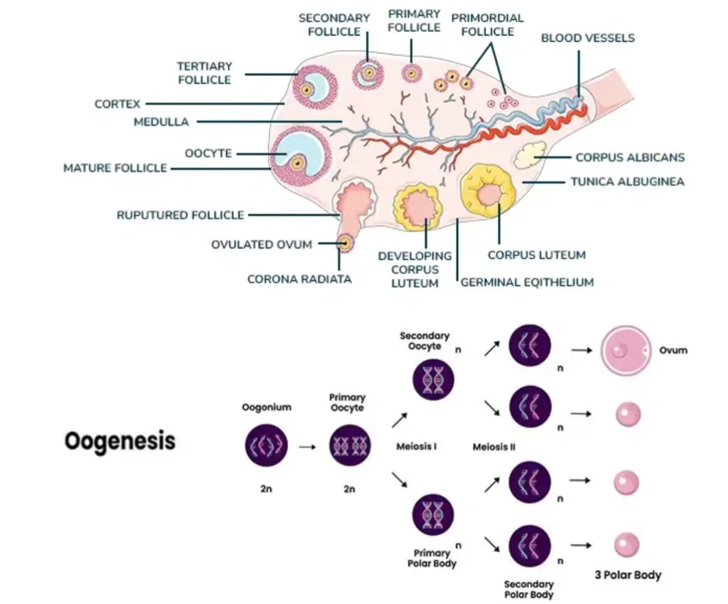
Oogenesis differs from spermatogenesis in that it begins long before birth:
- During foetal development, millions of gamete mother cells called oogonia form within the developing ovary.
- These oogonia enter meiosis but arrest at prophase I, becoming primary oocytes.
- Before puberty, a very large number of primary oocytes degenerate. The surviving primary oocytes become surrounded by layers of granulosa cells and a theca, forming secondary follicles.
- Secondary follicles develop a fluid-filled cavity called the antrum, becoming tertiary follicles. At this stage, the primary oocyte completes meiosis I, producing a haploid secondary oocyte and a small, non-functional first polar body.
- The tertiary follicle matures into the Graafian follicle, in which the secondary oocyte is surrounded by a protective glycoprotein layer called the zona pellucida.
- The Graafian follicle ruptures during ovulation, releasing the secondary oocyte from the ovary. (Meiosis II is only completed if the oocyte is fertilised.)
The Menstrual Cycle
The menstrual cycle is the reproductive cycle characteristic of humans and other primates. It begins at puberty — a first event known as menarche — and typically repeats every 28–29 days until menopause.
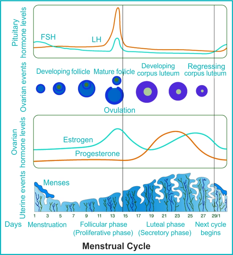
The menstrual cycle can be divided into three overlapping phases:
1. Menstrual Phase (Days 1–5)
- The cycle begins with menstrual flow, lasting about 3–5 days.
- This occurs because the endometrium breaks down and is shed, along with blood, if the released ovum was not fertilised in the previous cycle.
2. Follicular Phase (Days ~5–13)
- Primary follicles in the ovary grow and mature into Graafian follicles.
- Simultaneously, the endometrium regenerates through a proliferative phase.
- This phase is driven by rising levels of LH and FSH, which stimulate follicular growth. The growing follicles, in turn, secrete increasing amounts of oestrogen.
3. Ovulatory Phase (Around Day 14)
- LH and FSH levels peak mid-cycle (a surge often called the LH surge), triggering the rupture of the mature Graafian follicle.
- This releases the ovum from the ovary — the event known as ovulation.
4. Luteal Phase (Days ~15–28)
- The ruptured follicle transforms into the corpus luteum, a temporary endocrine structure.
- The corpus luteum secretes large amounts of progesterone, which is essential for maintaining the endometrium in preparation for a possible pregnancy.
- If fertilisation does not occur, the corpus luteum degenerates into a structure called the corpus albicans. Progesterone and oestrogen levels fall sharply, the endometrium breaks down, and a new menstrual cycle begins.
- If fertilisation does occur, the developing embryo (via hCG, discussed below) signals the corpus luteum to persist and continue secreting progesterone to sustain the pregnancy.
Quick fact: In humans, the menstrual cycle permanently stops around the age of 50 years — this stage is called menopause.
Fertilisation and Implantation
Fertilisation
- During coitus (sexual intercourse), semen is released (insemination) into the vagina. Motile sperm swim through the cervix and uterus and reach the ampullary-isthmic junction of the fallopian tube — the typical site of fertilisation.
- The ovum released during ovulation is also transported to this junction.
- Fertilisation is the fusion of a sperm and an ovum to form a zygote.
- When a sperm contacts the ovum, it triggers biochemical changes in the zona pellucida that block the entry of any additional sperm — this ensures only one sperm fertilises one ovum (a mechanism called the block to polyspermy).
- Enzymes released from the sperm’s acrosome help it penetrate the layers surrounding the ovum and enter its cytoplasm.
- Sperm entry triggers the secondary oocyte to complete its second meiotic division, producing a haploid ovum (ootid) and a second polar body.
- Finally, the haploid nucleus of the sperm fuses with the haploid nucleus of the ovum, restoring the diploid number of chromosomes and forming a zygote.
Cleavage and Implantation
- As the zygote travels down the oviduct toward the uterus, it undergoes rapid mitotic divisions called cleavage, producing progressively smaller cells called blastomeres (2 → 4 → 8 → 16 cells).
- The 8–16 celled embryo is known as a morula, which continues developing into a blastocyst as it moves into the uterus.
- Within the blastocyst, cells organise into two distinct groups:
- Trophoblast – the outer layer of cells
- Inner cell mass – a cluster of cells that will go on to form the embryo itself
- The trophoblast attaches to the uterine endometrium in a process called implantation — the moment that marks the true beginning of pregnancy.
- The inner cell mass subsequently differentiates to form the embryo proper.
Pregnancy, Placenta, and Foetal Development
- After implantation, the trophoblast develops finger-like projections called chorionic villi, which are surrounded by uterine tissue and maternal blood.
- The chorionic villi and adjoining uterine tissue together form the placenta — a specialised structure that supplies the growing embryo with oxygen and nutrients and removes metabolic waste.
- The placenta connects to the embryo through the umbilical cord.
- The placenta also functions as a temporary endocrine gland, secreting several essential hormones:
- Human Chorionic Gonadotropin (hCG) — this hormone is the basis of most home pregnancy tests
- Human Placental Lactogen (hPL)
- Oestrogens
- Progestogens
- Relaxin (secreted mainly in the later stages of pregnancy)
- These hormones — along with rising maternal levels of cortisol and prolactin — support foetal growth and help maintain the pregnancy.
- Shortly after implantation, the inner cell mass differentiates into three primary germ layers — the ectoderm, mesoderm, and endoderm — each of which gives rise to different tissues and organs. This developmental flexibility is possible because the inner cell mass contains multipotent stem cells.
Milestones of Foetal Development
| Timeline | Development |
| By end of 12 weeks | Most major organs of the body have formed |
| During the 5th month | Limbs and body hair develop |
| By the 24th week | Eyelids separate; eyelashes form |
| By the end of 9 months | The foetus is fully developed and ready for birth |
Parturition and Lactation
Parturition (Childbirth)
- Human pregnancy lasts approximately 9 months, a duration referred to as the gestation period.
- At the end of gestation, vigorous, rhythmic contractions of the uterus lead to the delivery of the foetus — this process is called parturition.
- Parturition is triggered by a neuroendocrine mechanism. Signals originating from the fully developed foetus and the placenta generate what is known as the foetal ejection reflex.
- This reflex stimulates the release of oxytocin from the mother’s pituitary gland.
- Oxytocin acts on the uterine muscle, causing increasingly strong contractions, which in a positive-feedback loop stimulate even more oxytocin release.
- These contractions ultimately lead to the expulsion of the baby, followed by the placenta (afterbirth).
Lactation
- During pregnancy, the mammary glands undergo extensive structural differentiation in preparation for milk production.
- Milk production begins around the time of childbirth.
- The very first milk secreted, produced during the first few days after delivery, is called colostrum. It is rich in antibodies (particularly IgA) and provides the newborn with vital passive immunity, helping protect it from infections during its earliest, most vulnerable days.
Quick Revision Summary
- Male gonad: Testes → produce sperm (spermatogenesis) and androgens
- Female gonad: Ovaries → produce ova (oogenesis) and oestrogen/progesterone
- Sperm pathway: Seminiferous tubules → rete testis → vasa efferentia → epididymis → vas deferens → ejaculatory duct → urethra
- Ovum pathway: Ovary → fimbriae → infundibulum → ampulla (fertilisation site) → isthmus → uterus
- Menstrual cycle length: 28–29 days on average
- Ovulation: occurs around Day 14 of the cycle
- Fertilisation site: ampullary-isthmic junction of the fallopian tube
- Implantation: trophoblast attaches to the endometrium → marks the start of pregnancy
- Gestation period: 9 months
- Birth-triggering hormone: oxytocin
- First milk: colostrum, rich in antibodies
Frequently Asked Questions (FAQs)
Q1. What are the main stages of human reproduction? The main stages are gametogenesis, insemination, fertilisation, implantation, gestation, and parturition.
Q2. Where does fertilisation occur in humans? Fertilisation takes place in the ampullary-isthmic junction of the fallopian tube (oviduct).
Q3. What is the difference between spermatogenesis and oogenesis? Spermatogenesis produces four functional haploid sperm from one primary spermatocyte and begins at puberty, continuing throughout adult life. Oogenesis produces only one functional haploid ovum (plus non-functional polar bodies) from one primary oocyte, and begins during foetal life, with oocytes remaining arrested until puberty and beyond.
Q4. What triggers parturition (labour)? Parturition is triggered by signals from the fully developed foetus and placenta known as the foetal ejection reflex, which stimulates the release of oxytocin, causing strong uterine contractions.
Q5. Why is colostrum important for a newborn? Colostrum, the first milk produced after childbirth, is rich in antibodies that help the newborn build immunity against infections in its early days of life.
These notes are designed for Class 12 Biology students following the NCERT curriculum and can also serve as a quick revision resource for competitive exams such as NEET.
Other topics you might be interested in: