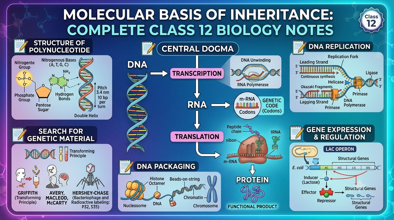
A complete, exam-focused guide to Chapter 6 of Class 12 Biology — covering DNA structure, replication, the central dogma, gene expression, the Human Genome Project, and DNA fingerprinting.
Introduction
Every trait you inherit — eye color, blood group, or a genetic disorder — is written in a molecule barely 2 nanometers wide: DNA. The chapter “Molecular Basis of Inheritance Class 12 Notes” explains how DNA stores genetic information, how it copies itself accurately, and how it directs the production of proteins that build and run our bodies.
This guide breaks the chapter into clear, structured sections designed for quick revision as well as deep understanding — ideal for CBSE, NEET, and other competitive exam preparation.
1. Structure of the Polynucleotide Chain
DNA (deoxyribonucleic acid) is a polymer built from repeating units called deoxyribonucleotides. Understanding its building blocks is the foundation of this entire chapter.
Nucleoside vs. Nucleotide
| Term | Composition | Bond Involved |
| Nucleoside | Nitrogenous base + Pentose sugar | N-glycosidic bond |
| Nucleotide | Nucleoside + Phosphate group | Phosphodiester bond |
- Examples of nucleosides: adenosine, deoxyadenosine, cytidine
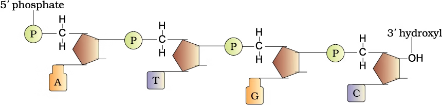
- Individual nucleotides link together through 3’–5′ phosphodiester bonds, forming a long polynucleotide chain (found in both DNA and RNA).
- Every polynucleotide chain has direction: a free phosphate group at the 5′ end and a free hydroxyl (–OH) group at the 3′ end.
2. The Double Helix Model of DNA
Key Scientists and Their Contributions
Understanding who discovered what is a frequently asked exam topic:
| Scientist(s) | Contribution |
| Friedrich Miescher | First identified DNA in the nucleus (1869); called it “Nuclein” |
| Maurice Wilkins & Rosalind Franklin | Produced the X-ray diffraction data that revealed DNA’s helical structure |
| James Watson & Francis Crick | Proposed the double helix model (1953) using Franklin’s X-ray data |
| Erwin Chargaff | Showed that in double-stranded DNA, A:T and G:C ratios are always equal to 1 (Chargaff’s Rule) |
Features of the DNA Double Helix
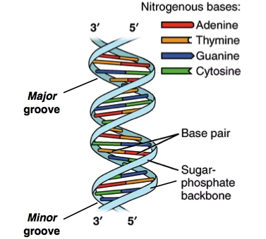
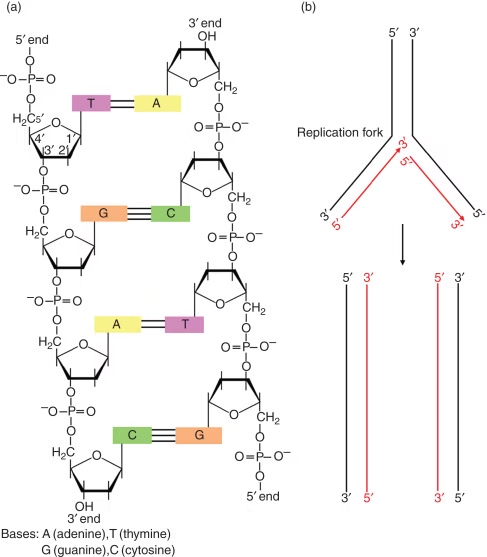
- DNA consists of two antiparallel polynucleotide chains coiled around a common axis to form a helix.
- The sugar-phosphate backbone lies on the outside, while the nitrogenous bases project inward.
- Bases pair through hydrogen bonding, and pairing always follows the purine–pyrimidine rule:
- Adenine (A) pairs with Thymine (T) — 2 hydrogen bonds
- Guanine (G) pairs with Cytosine (C) — 3 hydrogen bonds
- The helix is right-handed, with a pitch of 3.4 nm and 10 base pairs per turn.
- Base stacking (bases stacked one over another) adds further structural stability, in addition to hydrogen bonding.
3. Packaging of DNA in the Cell
A single human DNA molecule is astonishingly long — understanding how it fits inside a microscopic nucleus is a key concept.
The Scale of the Problem
- Distance between two consecutive base pairs = 0.34 nm (0.34 × 10⁻⁹ m)
- Total base pairs in human DNA ≈ 6.6 × 10⁹ bp
- Total length of human DNA ≈ 2.2 meters
- The nucleus, however, is only about 10⁻⁶ m in diameter — meaning DNA must be compacted roughly a million-fold.
DNA Organization in Prokaryotes
Prokaryotes lack a true nucleus. Their DNA is organized into large loops held in place by positively charged proteins, in a region called the nucleoid.
DNA Organization in Eukaryotes (Nucleosome Model)
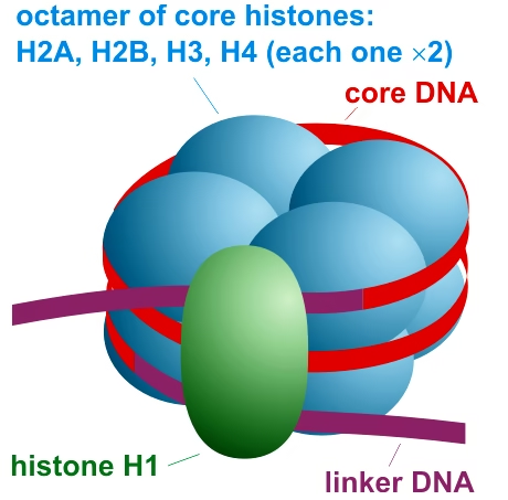
- Eukaryotic DNA is packaged with histones — small, positively charged basic proteins rich in the amino acids lysine and arginine.
- Eight histone molecules form a histone octamer.
- Negatively charged DNA winds around the positively charged histone octamer to form a nucleosome, which contains about 200 bp of DNA.
- A chain of nucleosomes looks like “beads on a string” under an electron microscope.
- These structures coil further into chromatin fibres, which condense further to form chromosomes during metaphase.
- Non-histone chromosomal proteins (NHC proteins) assist in this higher-order packaging.
Euchromatin vs. Heterochromatin
| Feature | Euchromatin | Heterochromatin |
| Packing | Loosely packed | Densely packed |
| Staining | Light | Dark |
| Transcriptional Activity | Active | Inactive |
4. The Search for the Genetic Material
Before DNA was confirmed as the genetic material, scientists debated whether it was DNA or protein. Three landmark experiments settled the question.
Griffith’s Transformation Experiment (1928)
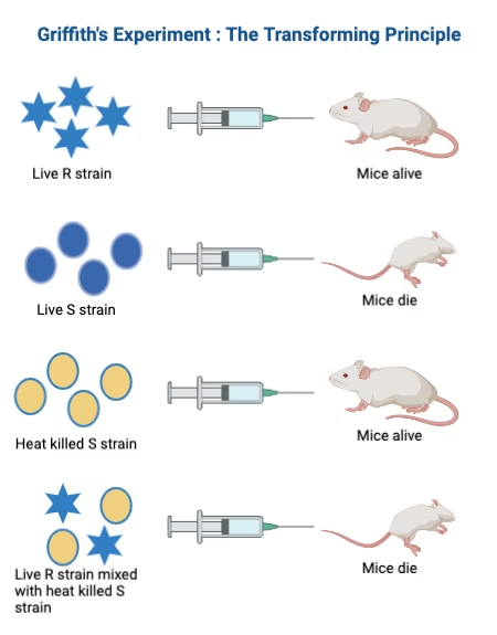
Frederick Griffith worked with two strains of Streptococcus pneumoniae:
| S Strain (Smooth) | R Strain (Rough) |
| Has a polysaccharide coat | No polysaccharide coat |
| Virulent — causes pneumonia | Non-virulent — harmless |
Griffith’s key observations:
- Live S strain → mouse dies
- Live R strain → mouse survives
- Heat-killed S strain → mouse survives
- Heat-killed S strain + Live R strain → mouse dies
In the fourth case, the dead mouse’s blood contained living S strain bacteria. Griffith concluded that some “transforming principle” from the dead S bacteria had transferred genetic information to the harmless R bacteria, converting it into a virulent form. This phenomenon is known as transformation.
Avery, MacLeod & McCarty: Identifying the Transforming Principle
To find out which biomolecule was responsible for transformation, these scientists treated the heat-killed S strain + R strain mixture with different enzymes:
- Treating with proteases and RNase (which destroy proteins and RNA) → transformation still occurred
- Treating with DNase (which destroys DNA) → transformation was blocked
This proved that DNA — not protein or RNA — is the genetic material.
The Hershey-Chase Experiment (1952)
Alfred Hershey and Martha Chase provided the final, definitive proof using bacteriophages (viruses that infect bacteria):
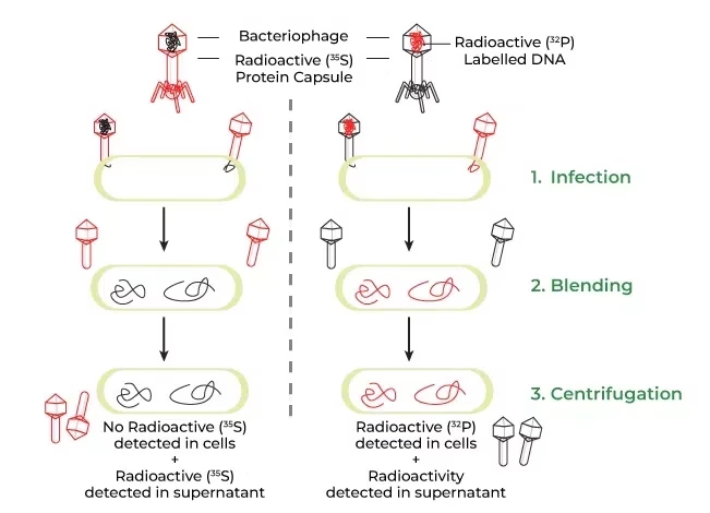
- One batch of phages was labeled with radioactive sulphur (³⁵S), which tags proteins (since sulphur is absent in DNA).
- Another batch was labeled with radioactive phosphorus (³²P), which tags DNA (since phosphorus is absent in most amino acids).
- Both batches were used to infect E. coli, then the mixture was blended and centrifuged to separate viral coats from bacterial cells.
Results:
- Bacteria infected with ³²P-labeled phages were radioactive → DNA had entered the cells.
- Bacteria infected with ³⁵S-labeled phages were not radioactive → protein stayed outside.
Conclusion: DNA, not protein, is the genetic material that is passed from virus to host.
5. Properties of Genetic Material: Why DNA (and not RNA)?
For any molecule to serve as effective genetic material, it must:
- Be able to replicate accurately.
- Be chemically and structurally stable.
- Allow for occasional mutations, providing the raw material for evolution.
- Be capable of expression as observable traits (following Mendelian inheritance).
DNA vs. RNA
| Feature | DNA | RNA |
| Sugar | Deoxyribose | Ribose |
| Base | Thymine | Uracil |
| Strand | Usually double-stranded | Usually single-stranded |
| Stability | Highly stable | Less stable, easily degraded |
| Role | Primary genetic material | Messenger, adaptor; genetic material only in some viruses |
Why is DNA more stable than RNA?
- RNA has a reactive 2′-OH group on every nucleotide, making it chemically unstable and prone to degradation.
- DNA contains thymine instead of uracil, which adds chemical stability.
- Because DNA is double-stranded, any damage on one strand can be repaired using the complementary strand as a template — a self-correcting mechanism unavailable to single-stranded RNA.
This is why DNA, being more stable, evolved as the primary long-term storage molecule for genetic information, while the more reactive RNA plays roles in gene expression.
6. DNA Replication
DNA replication is the process by which a cell produces an identical copy of its DNA before cell division.
Semi-Conservative Replication
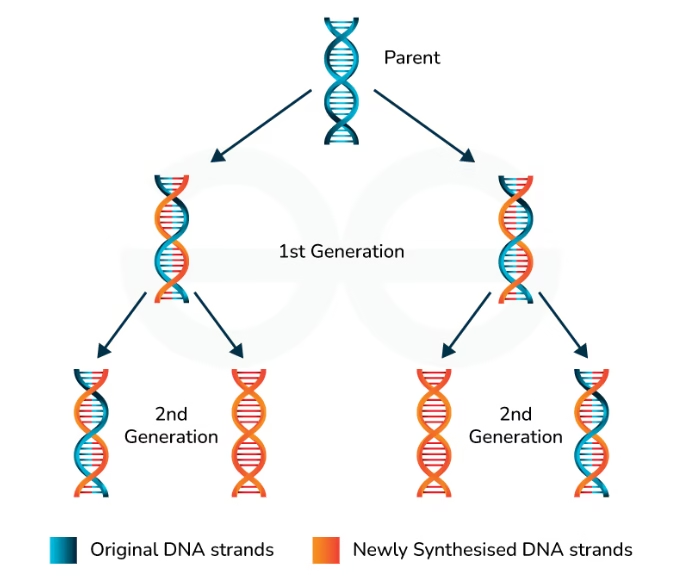
Watson and Crick’s structure itself suggested a mechanism: each strand separates and acts as a template for synthesizing a new complementary strand. The result is two DNA molecules, each with one parental (old) strand and one newly synthesised strand — this is called semi-conservative replication.
The Meselson-Stahl Experiment (Proof of Semi-Conservative Replication)
Matthew Meselson and Franklin Stahl confirmed this model using E. coli and isotopes of nitrogen:
- E. coli was grown in a medium with the heavy isotope ¹⁵N, producing “heavy” DNA.
- Cells were then shifted to a medium with normal ¹⁴N.
- DNA samples were extracted at intervals and separated using cesium chloride density gradient centrifugation.
Observations:
- After one generation (20 minutes): all DNA showed a hybrid density (one ¹⁵N strand + one ¹⁴N strand).
- After two generations (40 minutes): DNA showed equal amounts of hybrid and light (fully ¹⁴N) density.
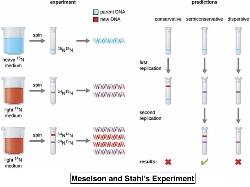
This pattern is only possible if replication is semi-conservative, confirming Watson and Crick’s prediction.
Mechanism of Replication
- Replication occurs during the S phase of the cell cycle.
- Enzyme: DNA polymerase (a DNA-dependent DNA polymerase).
- Energy source: Deoxyribonucleoside triphosphates (dNTPs), which serve the dual role of substrate and energy supply.
- Replication starts at a specific site called the origin of replication.
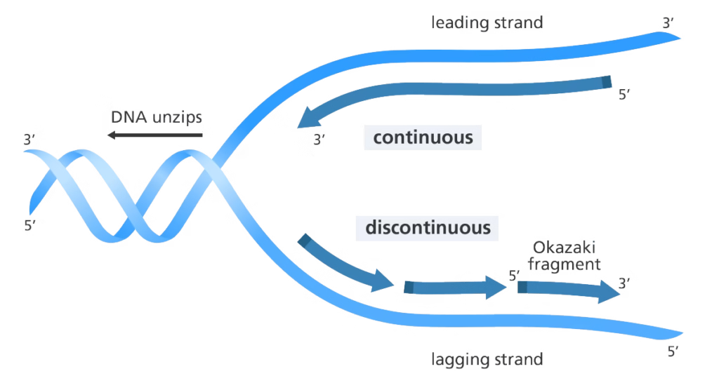
- The DNA double helix opens locally to form a replication fork.
- DNA polymerase synthesizes new strands only in the 5’→3′ direction:
- On one template strand, synthesis is continuous (leading strand).
- On the other, synthesis is discontinuous, producing short fragments called Okazaki fragments (lagging strand).
- These fragments are later joined together by the enzyme DNA ligase.
7. Transcription: DNA to RNA
Transcription is the process of copying genetic information from DNA into RNA.
Why Only One Strand Is Copied
If both DNA strands were transcribed simultaneously:
- They would produce two different RNA sequences, coding for two unrelated proteins from a single gene.
- The two RNA molecules, being complementary to each other, would immediately pair up to form double-stranded RNA — making translation impossible and the entire process pointless.
Hence, only a specific segment of one strand is used as the template.
Structure of a Transcription Unit
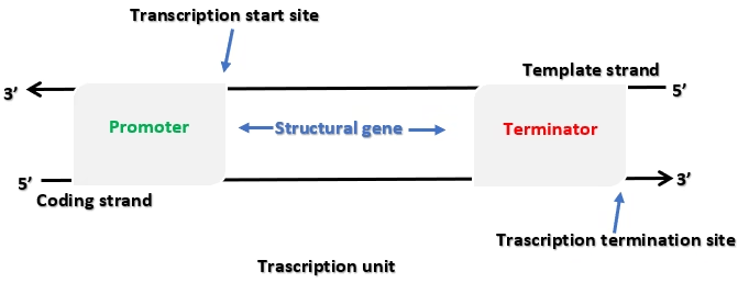
A transcription unit has three components:
- Promoter — Signals the start of transcription; the site where RNA polymerase binds.
- Structural gene — The actual DNA sequence being transcribed.
- Terminator — Marks the end of transcription.
Template Strand vs. Coding Strand
- The strand read by RNA polymerase (in the 3’→5′ direction) is the template strand.
- The other strand, with the same sequence as the RNA transcript (except T is replaced by U), is called the coding strand — though technically it doesn’t “code” for anything directly.
The Three Stages of Transcription
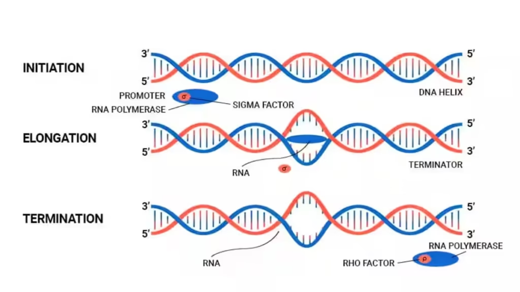
- Initiation: RNA polymerase binds to the promoter with the help of an initiation factor (sigma, σ), which increases its binding specificity.
- Elongation: RNA polymerase moves along the template, adding ribonucleotides complementary to the DNA template.
- Termination: A termination factor (rho, ρ) signals RNA polymerase to release the newly formed RNA transcript.
Types of RNA and Their Roles
| RNA Type | Function |
| mRNA (messenger RNA) | Carries the genetic code from DNA to the ribosome for protein synthesis |
| tRNA (transfer RNA) | Reads the genetic code and delivers the corresponding amino acid |
| rRNA (ribosomal RNA) | Structural and catalytic component of the ribosome |
Transcription in Eukaryotes vs. Prokaryotes
Prokaryotes: Since there is no nuclear membrane, transcription and translation occur simultaneously (coupled) — translation can begin before transcription is even complete.
Eukaryotes: Transcription is more complex, involving three distinct RNA polymerases:
| Enzyme | Function |
| RNA Polymerase I | Transcribes rRNA |
| RNA Polymerase II | Transcribes hnRNA (the mRNA precursor) |
| RNA Polymerase III | Transcribes tRNA, snRNA, and other small RNAs |
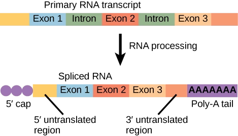
The primary transcript, hnRNA (heterogeneous nuclear RNA), undergoes three processing steps before it becomes mature mRNA:
- Splicing — Removal of non-coding introns and joining of coding exons.
- Capping — Addition of methyl guanosine triphosphate to the 5′ end.
- Tailing — Addition of a poly-A tail (adenylate residues) to the 3′ end.
Only after these modifications does hnRNA become mature mRNA, which is exported from the nucleus for translation.
What Is a Gene?
- A gene is the DNA sequence that codes for one polypeptide, tRNA, or rRNA molecule.
- A cistron is a segment of DNA coding for one polypeptide.
- Genes can be:
- Monocistronic — common in eukaryotes, containing exons and introns
- Polycistronic — common in prokaryotes
8. The Genetic Code
The genetic code defines how the sequence of nucleotides in mRNA determines the sequence of amino acids in a protein.
Cracking the Code
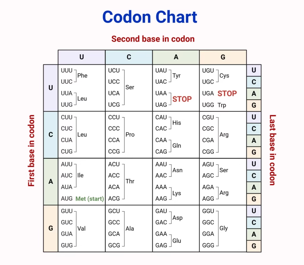
- George Gamow proposed that since only 4 bases must code for 20 amino acids, the code had to be at least a triplet (4³ = 64 combinations — more than enough, while 4² = 16 is insufficient).
- Har Gobind Khorana developed chemical methods to synthesize RNA molecules with defined, known sequences.
- Marshall Nirenberg developed cell-free protein-synthesizing systems that helped decode which codon specifies which amino acid.
- The enzyme polynucleotide phosphorylase (identified through the work of Severo Ochoa) helped polymerize RNA of a random, defined sequence, further aiding decoding efforts.
Salient Features of the Genetic Code
- Triplet code: Each codon consists of 3 nucleotides. Of the 64 possible codons, 61 code for amino acids and 3 are stop codons.
- Unambiguous: Each codon specifies only one particular amino acid.
- Degenerate: Multiple codons can code for the same amino acid.
- Universal: The same codon specifies the same amino acid across nearly all living organisms — a strong piece of evidence for a common evolutionary origin of life.
- Non-overlapping and comma-less: Codons are read continuously in one direction without gaps.
- AUG has a dual role: It codes for methionine and also serves as the start codon.
Mutations and the Reading Frame
- Point mutations — a single base change — can alter one amino acid, sometimes with drastic effects. A classic example is sickle cell anaemia, caused by a change from Glutamic acid to Valine in the beta-globin chain of hemoglobin.
- Frameshift mutations occur when a base is inserted or deleted, shifting the entire reading frame downstream.
- Interestingly, inserting or deleting three (or a multiple of three) bases together restores the original reading frame — a fact historically used as key evidence that the codon is indeed a triplet.
9. tRNA Structure and Translation
Structure of tRNA
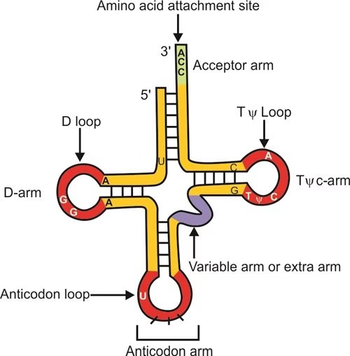
- tRNA is the adapter molecule of protein synthesis — it reads the genetic code on one end and carries a specific amino acid on the other.
- Its anticodon loop pairs with the complementary codon on mRNA.
- Its amino acid acceptor end binds to the specific amino acid corresponding to that codon.
- A special initiator tRNA carries methionine and recognizes the AUG start codon.
- No tRNA exists for the three stop codons.
Steps of Translation
- Charging (aminoacylation) of tRNA: Amino acids are activated using ATP and attached to their specific tRNA.
- Initiation: The small ribosomal subunit binds to mRNA; the initiator tRNA recognizes the start codon (AUG).
- Elongation: Charged tRNAs bind to successive codons through complementary base pairing between codon and anticodon. Amino acids are joined via peptide bonds, and the ribosome moves codon by codon along the mRNA.
- Termination: When the ribosome reaches a stop codon, a release factor binds, releasing the completed polypeptide chain.
Additional key terms:
- Translational unit: The region of mRNA between the start codon and the stop codon.
- UTR (Untranslated Region): Non-coding sequences before the start codon (5′ UTR) or after the stop codon (3′ UTR) that assist translation efficiency without being translated themselves.
10. Regulation of Gene Expression: The Lac Operon
Cells don’t waste energy making proteins they don’t need. Gene regulation ensures genes are switched on only when required — the lac operon in E. coli is the textbook model of this process.
Levels of Regulation
Gene expression can be controlled at multiple stages:
- Transcriptional level
- RNA processing (splicing)
- Transport of mRNA from nucleus to cytoplasm
- Translational level
The Lac Operon: Structure and Genes
Discovered by Francois Jacob and Jacques Monod, the lac operon is a polycistronic gene cluster regulated by a shared promoter and operator.
| Gene | Type | Function |
| i gene | Regulatory | Codes for the repressor protein |
| z gene | Structural | Codes for β-galactosidase (breaks lactose into glucose + galactose) |
| y gene | Structural | Codes for permease (increases membrane permeability to lactose) |
| a gene | Structural | Codes for transacetylase |
How the Lac Operon Works
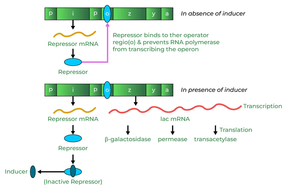
In the absence of lactose (inducer):
- The i gene continuously produces a repressor protein.
- The repressor binds tightly to the operator region, physically blocking RNA polymerase from transcribing the z, y, and a genes.
- Result: lactose-metabolizing enzymes are not produced.
In the presence of lactose (inducer):
- Lactose (or its derivative) binds to the repressor protein, inactivating it.
- The inactive repressor can no longer bind the operator.
- RNA polymerase is now free to transcribe z, y, and a, producing β-galactosidase, permease, and transacetylase.
- These enzymes enable the cell to metabolize lactose efficiently.
This elegant feedback system ensures the lac operon is only “switched on” when its substrate — lactose — is actually available, conserving cellular energy.
11. The Human Genome Project (HGP)
Overview
- A collaborative venture between the US Department of Energy and the National Institutes of Health (NIH), later joined by the Wellcome Trust (UK).
- Launched in 1990, completed in 2003.
- Goal: to determine the complete sequence of base pairs that make up human DNA and identify all human genes.
Why It Mattered: Birth of Bioinformatics
- The human genome contains roughly 3 × 10⁹ base pairs.
- At an estimated cost of $3 per base pair, sequencing the entire genome would have cost around $9 billion.
- The resulting data — equivalent to about 3,300 books of 1,000 pages each — was too vast to manage manually, giving rise to the field of bioinformatics for storing, retrieving, and analyzing genomic data.
- Alongside humans, the genomes of several model organisms — bacteria, yeast, Caenorhabditis elegans, Drosophila, rice, and Arabidopsis — have also been sequenced.
Methods Used to Identify Genes
- Expressed Sequence Tags (ESTs): Sequencing only the parts of DNA that are actually transcribed into mRNA.
- Sequence Annotation: Sequencing the entire genome (coding and non-coding regions) and later assigning functions to specific regions.
The Genome Sequencing Process
- DNA is isolated from cells and randomly fragmented.
- Fragments are cloned into vectors such as BAC (Bacterial Artificial Chromosomes) and YAC (Yeast Artificial Chromosomes) and inserted into host cells (bacteria/yeast) for amplification.
- Amplified fragments are sequenced using automated Sanger sequencing methods.
- Sequences are aligned based on overlapping regions using computer software.
- Finally, sequences are annotated and mapped to specific chromosomes.
Genetic and physical maps of the genome are also built using restriction fragment length polymorphism (RFLP) and microsatellite (repetitive DNA) analysis.
Key Findings from the Human Genome Project
- The human genome contains approximately 3,164.7 million nucleotide bases.
- An average gene is about 3,000 bases long, though sizes vary widely — the largest known gene, dystrophin, spans 2.4 million bases.
- The human genome contains an estimated 30,000 genes — far fewer than scientists originally expected.
- Functions of over 50% of discovered genes remain unknown.
- Less than 2% of the genome actually codes for proteins.
- A large fraction of the genome consists of repetitive, non-coding sequences, which may play roles in evolution and chromosome structure.
- Chromosome 1 has the most genes (2,968); Chromosome Y has the fewest (231).
- Single Nucleotide Polymorphisms (SNPs) occur at roughly 1.4 million locations across the human genome and are important markers for studying disease and human evolutionary history.
12. DNA Fingerprinting
What Is DNA Fingerprinting?
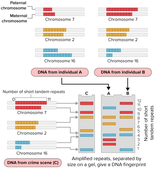
DNA fingerprinting is a technique used to compare specific regions of DNA between individuals, based on the fact that while 99.9% of human DNA sequences are identical across individuals, the remaining 0.1% makes each person genetically unique (except identical twins).
Rather than sequencing all 3 billion base pairs — an impractical task — scientists focus on highly variable repetitive DNA sequences.
The Science Behind It
- When genomic DNA is separated by density gradient centrifugation, most of it forms a large “bulk DNA” peak, while a smaller fraction forms distinct peaks known as satellite DNA.
- Satellite DNA is classified into microsatellites and minisatellites based on base composition, segment length, and repeat number.
- Though satellite DNA doesn’t code for proteins, it shows a high degree of polymorphism — variation between individuals — making it ideal for identification purposes.
- This polymorphism arises from mutations in germ cells, which get passed down through generations. Because non-coding regions don’t affect reproductive fitness, mutations accumulate there more freely.
Methodology
DNA fingerprinting relies on VNTRs (Variable Number of Tandem Repeats) — a class of satellite DNA showing high polymorphism, used as probes.
Step-by-step process:
- DNA is isolated from the individual and cut using restriction endonucleases.
- The resulting fragments are separated by size using gel electrophoresis.
- Separated fragments are transferred (blotted) onto a nylon or nitrocellulose membrane.
- The membrane is hybridized with a labeled VNTR probe.
- Hybridized fragments are visualized through autoradiography, revealing a unique pattern of bands.
Since VNTRs range from 0.1 to 20 kb in size, the resulting band pattern is highly individual-specific — no two people (except identical twins) show the exact same pattern.
Applications of DNA Fingerprinting
- Forensic science: Identifying suspects or victims from biological evidence, since every tissue from one individual shows the same degree of polymorphism.
- Paternity testing: A child inherits half its polymorphic pattern from each parent, allowing biological parentage to be confirmed.
- Evolutionary and population studies: Assessing genetic diversity within and between populations.
Quick Revision Summary
| Concept | Key Fact |
| DNA structure | Double helix, antiparallel strands, A-T (2 H-bonds), G-C (3 H-bonds) |
| Genetic material proof | Griffith → Avery-MacLeod-McCarty → Hershey-Chase |
| Replication | Semi-conservative (Meselson-Stahl); enzyme: DNA polymerase |
| Transcription | DNA → RNA; enzyme: RNA polymerase; only one strand copied |
| Genetic code | Triplet, degenerate, unambiguous, universal; 61 sense + 3 stop codons |
| Translation | mRNA → protein; occurs on ribosomes with tRNA as adapter |
| Gene regulation | Lac operon — inducible, negative regulation by repressor |
| HGP | 1990–2003; ~30,000 human genes; <2% genome coding |
| DNA fingerprinting | Based on VNTR polymorphism; used in forensics & paternity testing |
Frequently Asked Questions (FAQs)
Q1. What is the difference between a nucleoside and a nucleotide? A nucleoside is simply a nitrogenous base attached to a sugar. A nucleotide is a nucleoside plus a phosphate group, making it the true building block of DNA and RNA.
Q2. Why did Hershey and Chase use radioactive sulphur and phosphorus? Because sulphur is present only in proteins (not DNA) and phosphorus is present only in DNA (not most proteins), these isotopes allowed the scientists to track exactly which molecule — protein or DNA — physically entered the bacterial cell during viral infection.
Q3. Why is DNA replication called semi-conservative? Because each of the two daughter DNA molecules retains one original (parental) strand and gains one newly synthesized strand — the genetic material is “conserved” in a half-and-half manner rather than being fully copied or fully retained.
Q4. What is the difference between a template strand and a coding strand? The template strand is physically read by RNA polymerase during transcription. The coding strand is not read directly but has the same sequence as the resulting mRNA (except thymine is replaced by uracil).
Q5. Why is the genetic code considered “universal”? Because the same codon specifies the same amino acid in almost all organisms, from bacteria to humans — strong evidence that all life shares a common evolutionary ancestor.
Q6. What is the main achievement of the Human Genome Project? It successfully mapped and sequenced nearly all of the roughly 3 billion base pairs in human DNA, identifying about 30,000 genes and laying the foundation for modern genomics and personalized medicine.
These notes are designed for Class 12 Biology (CBSE/NCERT-aligned) students preparing for board exams and competitive exams such as NEET. For best results, pair this reading with diagram practice (DNA structure, replication fork, lac operon) and previous years’ question papers.
Other topics you might be interested in:
Class 12 Biology Notes: Reproduction in Organisms
Sexual Reproduction in Flowering Plants: Class 12 Biology Notes
Human Reproduction Class 12 Notes Biology
Principles of Inheritance and Variation — Class 12 Biology Notes