Agarose Gel Electrophoresis is one of the most widely used techniques in molecular biology, genetics, and biochemistry. It plays a central role in DNA and RNA analysis, genetic fingerprinting, and even in medical diagnostics. This method allows scientists to separate and visualize nucleic acids (DNA and RNA) based on their size and charge.
In this article, we will learn in detail about the principle, parts of the apparatus, step-by-step procedure, concentration of agarose gel, applications, advantages, disadvantages, and frequently asked questions.
Introduction to Agarose Gel Electrophoresis
- Definition: Agarose Gel Electrophoresis is a laboratory technique used to separate DNA, RNA, or proteins in a gel-like medium called agarose gel, using an electric current.
- Nature of agarose: Agarose is a natural polysaccharide (a type of sugar polymer) obtained from seaweed. When dissolved in buffer and cooled, it forms a gel matrix with tiny pores. These pores act like a sieve, allowing smaller DNA fragments to move faster than larger ones.
- Why it is important: It is one of the simplest, cheapest, and most effective techniques for analyzing nucleic acids. Students, researchers, and clinical scientists regularly use it to check DNA quality, run PCR products, or study gene expression.
Principle of Agarose Gel Electrophoresis
The technique works on two main principles:
- Charge of DNA/RNA
- DNA and RNA molecules have a negatively charged backbone due to phosphate groups.
- When an electric field is applied, these molecules move towards the positive (anode) electrode.
- Size-based separation
- Smaller fragments of DNA move faster through the pores of agarose gel.
- Larger fragments face more resistance and move slower.
- This allows separation of DNA fragments of different lengths.
- Visualization of DNA
- To see DNA, dyes like Ethidium Bromide (EtBr) or safer alternatives (e.g., SYBR Green, GelRed) are used.
- These dyes bind to DNA and fluoresce when exposed to UV light, making DNA bands visible.
*Key point: Smaller DNA fragments are found near the bottom of the gel, while larger fragments remain closer to the wells at the top.
Components and Requirements of Agarose Gel Electrophoresis
To perform this experiment, the following instruments and materials are required:
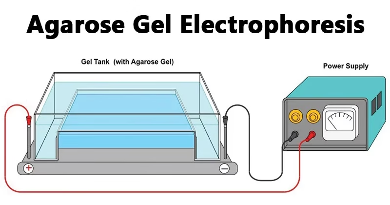
- Electrophoresis chamber and power supply
- Provides a controlled electric field for the movement of DNA fragments.
- Gel casting tray
- A plastic tray (UV transparent) used to prepare the agarose gel.
- Sample combs
- Small comb-like structures placed in the gel before solidification to create wells where DNA samples are loaded.
- Electrophoresis buffer
- Maintains pH and provides ions for conducting electricity.
- Common buffers: TAE (Tris-Acetate-EDTA) and TBE (Tris-Borate-EDTA).
- Loading buffer
- Mixed with DNA samples before loading.
- Contains:
- Glycerol (to make the sample heavier so it sinks into wells).
- Tracking dyes (e.g., Bromophenol blue, Xylene cyanol) to monitor DNA migration.
- Staining agents
- Ethidium Bromide (EtBr) – most common but carcinogenic.
- Alternatives: SYBR Green, GelRed (safer).
- Transilluminator (UV light box)
- Used to visualize DNA bands after electrophoresis.
Steps in Agarose Gel Electrophoresis
The process can be divided into several key steps:
Step 1: Preparation of Gel
- Weigh agarose powder and dissolve it in buffer (TAE/TBE).
- Heat the mixture in a microwave until agarose dissolves completely.
- Cool the solution to ~60°C and pour into casting tray with comb in place.
- Let it solidify at room temperature.
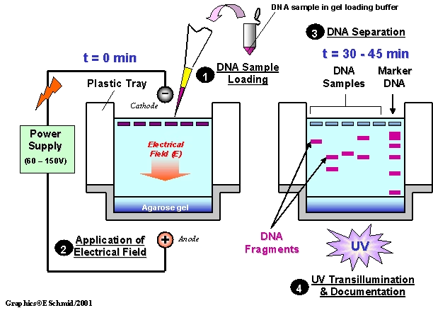
Step 2: Loading Samples
- Place the solidified gel into the electrophoresis chamber and cover it with buffer.
- Carefully remove the comb to create wells.
- Mix DNA sample with loading buffer and pipette into wells.
Step 3: Running the Gel
- Attach the chamber to a power supply.
- DNA migrates towards the positive electrode (red end).
- Monitor progress using tracking dyes.
Step 4: Visualization of DNA
- After electrophoresis, observe DNA bands under UV light.
- Compare with a DNA ladder/marker (fragments of known size) to estimate sizes of unknown DNA.
Concentration of Agarose Gel
The pore size of agarose gel depends on its concentration.
- Low concentration gel (0.2–0.7%) → large pores → useful for separating large DNA fragments.
- Medium concentration gel (0.8–1.5%) → commonly used for general DNA analysis (PCR products, plasmids).
- High concentration gel (2–3%) → small pores → useful for separating small DNA fragments.
Example:
- To separate DNA fragments of 100–1000 base pairs, a 2% agarose gel is used.
- To separate 10,000 bp or higher, a 0.7% agarose gel is recommended.
Choosing the Right Agarose Concentration
| Agarose Concentration (%) | Effective Range of Separation (kb) | Best For |
| 0.5% | 1 – 30 kb | Very large DNA fragments, genomic DNA |
| 0.7% | 0.8 – 12 kb | Routine analysis, Southern blotting, large PCR products |
| 1.0% | 0.5 – 10 kb | General purpose, most common concentration |
| 1.2% | 0.4 – 7 kb | Typical PCR products, restriction digests |
| 1.5% | 0.2 – 3 kb | Small PCR products, small fragments |
| 2.0% | 0.1 – 2 kb | Very small DNA fragments, <500 bp |
| 3.0% | 0.05 – 1 kb | Tiny fragments, like microsatellites |
Applications of Agarose Gel Electrophoresis
This technique has numerous applications in research, medicine, and diagnostics:
- DNA Fragment Analysis
- Estimation of DNA size using a marker or ladder.
- PCR Product Verification
- To check if amplification was successful in molecular biology experiments.
- Restriction Fragment Analysis
- Used in cloning and restriction mapping.
- Genetic Fingerprinting
- Used in forensic science for criminal identification.
- Medical Diagnostics
- Detection of genetic diseases by analyzing patient DNA.
- RNA Analysis
- Separation of RNA molecules for gene expression studies.
- Purification of DNA Fragments
- DNA bands can be cut out of gels for cloning experiments.
Advantages of Agarose Gel Electrophoresis
- Simple and easy to perform.
- Inexpensive compared to other techniques.
- Does not denature DNA or RNA.
- Samples can be recovered from gel.
- Suitable for a wide range of DNA fragment sizes.
Disadvantages of Agarose Gel Electrophoresis
- Gels can melt if voltage/current is too high.
- Buffers can become exhausted after long runs.
- Different DNA structures (linear, circular, supercoiled) migrate unpredictably.
- Limited resolution compared to polyacrylamide gels.
Safety Considerations
- Ethidium Bromide is toxic and mutagenic → always wear gloves and dispose waste properly.
- Use safer dyes like SYBR Safe whenever possible.
- UV light exposure can damage skin and eyes → use protective shields.
FAQs on Agarose Gel Electrophoresis
Q1. Why is agarose gel electrophoresis preferred for DNA separation?
Because agarose gels are easy to prepare, cheap, and suitable for separating large DNA fragments (up to 25 kb).
Q2. What is the difference between TAE and TBE buffer?
TAE has lower buffering capacity but allows faster migration, while TBE provides better resolution for small DNA fragments.
Q3. Can agarose gel electrophoresis separate proteins?
Yes, but it is not commonly used. Proteins are usually separated using SDS-PAGE.
Q4. What is a DNA ladder?
👉 A mixture of DNA fragments of known sizes, used as a reference to estimate the size of unknown DNA samples.
Q5. How long does agarose gel electrophoresis take?
Usually 30–60 minutes, depending on gel concentration and fragment size.
Conclusion
Agarose Gel Electrophoresis is an indispensable technique in modern biology and medicine. From basic DNA analysis in classrooms to advanced applications in genetics, forensics, and diagnostics, this simple yet powerful method continues to play a vital role in understanding the molecular basis of life.
By mastering the principles, steps, and applications of this technique, students and researchers gain a strong foundation in molecular biology experiments.
References
- https://pdfs.semanticscholar.org/36cf/d4ada922c44d233b6ebfa2af2c956c92e4ec.pdf
- https://www.mun.ca/biology/scarr/Gel_Electrophoresis.html
- https://www.wou.edu/las/physci/ch462/Gel%20Electrophoresis.pdf
- https://en.wikipedia.org/wiki/Agarose_gel_electrophoresis
- https://msu.edu/course/css/451/Lecture/PT-electrophoresis%20(2009).pdf
- http://library.umac.mo/ebooks/b28050459.pdf