Introduction to Gel Electrophoresis
- Electrophoresis is a widely used laboratory technique in biology, chemistry, medicine, and forensic science. It refers to the movement of charged molecules in an electric field.
- The method was pioneered in the 1930s by Arne Tiselius, a Swedish biophysicist, who studied the movement of proteins in an electric field. His contribution earned him the 1948 Nobel Prize in Chemistry.
- Over the years, electrophoresis evolved into different forms, with gel electrophoresis being the most popular and powerful version.
- Gel electrophoresis is mainly used to separate DNA, RNA, and proteins based on their size, charge, and shape. It is one of the cornerstones of molecular biology and biotechnology.
In simple words: Gel electrophoresis is like a molecular sieve that helps scientists “see” and separate biomolecules.
Principle of Gel Electrophoresis
- Most biomolecules (DNA, RNA, proteins) carry electrical charges because of their ionizable groups.
- When placed in an electric field, these charged molecules migrate:
- Negatively charged molecules (e.g., DNA, RNA) move towards the positive electrode (anode).
- Positively charged molecules move towards the negative electrode (cathode).
- The speed of migration depends on:
- Size (smaller molecules move faster).
- Charge (more charged molecules move faster).
- Shape (linear vs folded).
- Gel pore size.
This differential movement allows separation and identification of biomolecules.
Parts of a Gel Electrophoresis Apparatus
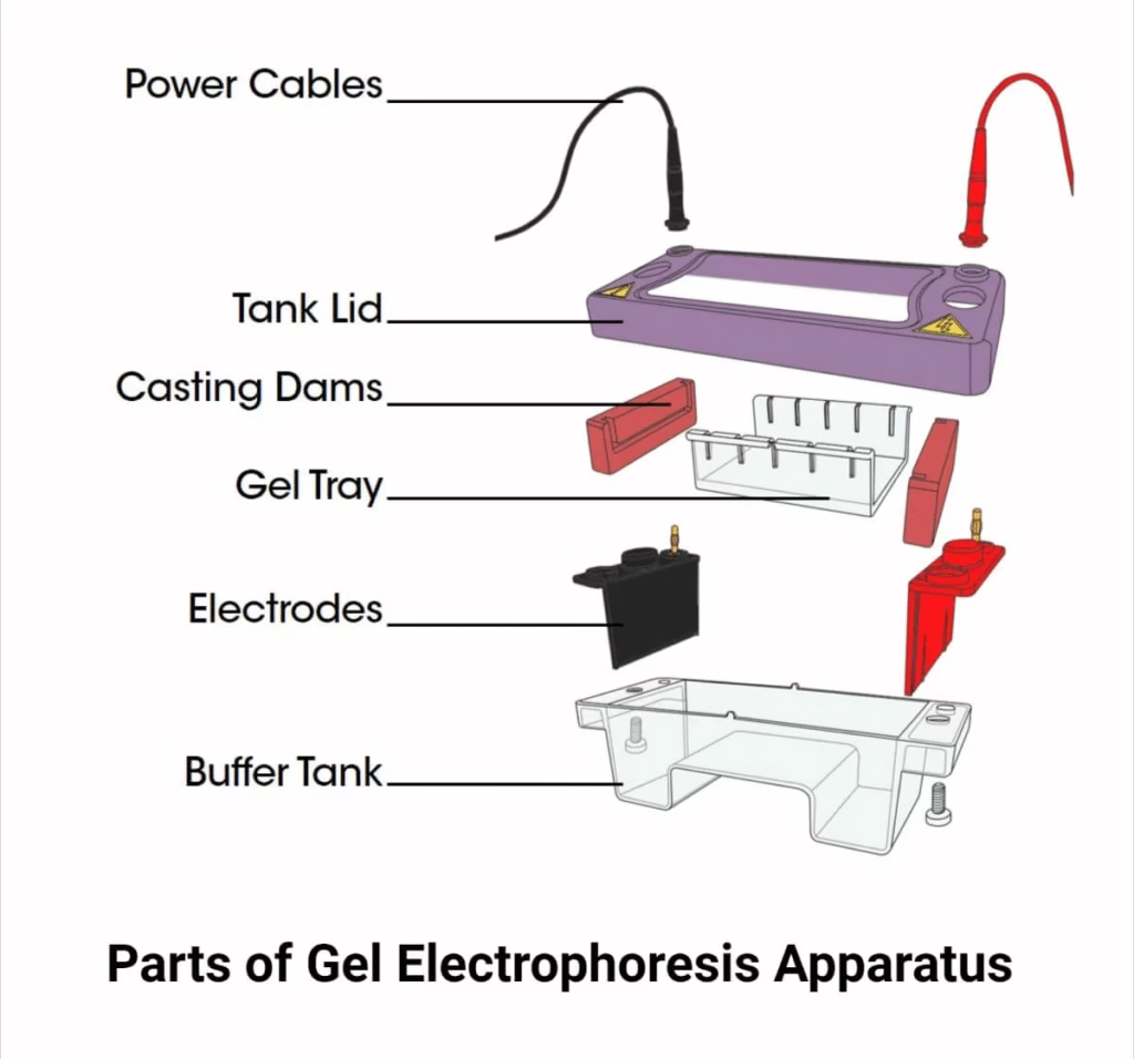
A typical gel electrophoresis setup has several components:
1. Power Supply
- Provides a constant voltage/current for molecule migration.
- Connected with red (anode, +) and black (cathode, –) cables.
- Controls the speed and resolution of separation.
2. Buffers
- Maintain pH and provide ions for current conduction.
- Common buffers: TAE (Tris-acetate-EDTA), TBE (Tris-borate-EDTA), Tris-glycine.
- Buffers also prevent overheating and sample degradation.
3. Supporting Media (Gel Matrix)
- Serves as a molecular sieve for separation.
- Types:
- Starch Gel – early medium, now obsolete.
- Cellulose Acetate – for protein electrophoresis.
- Agarose Gel – ideal for DNA/RNA (0.8–5%).
- Polyacrylamide Gel (PAGE) – for proteins and small DNA fragments.
4. Electrophoresis Chamber (Gel Box)
- Plastic tank filled with buffer.
- Holds gel slab, electrodes, and lid.
5. Electrodes
- Platinum or stainless steel.
- Attract oppositely charged molecules.
6. Gel Caster and Comb
- Used to prepare gel and wells where samples are loaded.
7. Staining and Destaining Containers
- For visualizing biomolecules after separation.
- Common stains: Ethidium bromide (EtBr), SYBR Green, Coomassie Brilliant Blue, Silver stain.
Types of Gel Electrophoresis
Gel electrophoresis comes in many forms depending on application:
1. Paper Gel Electrophoresis
- Early technique using filter paper as medium.
- Used for serum proteins.
- Disadvantages: poor resolution, protein adsorption.
2. Agarose Gel Electrophoresis
- Most common for DNA and RNA.
- Separation range: 100 bp – 20 kb DNA fragments.
- Pore size depends on agarose concentration.
3. Polyacrylamide Gel Electrophoresis (PAGE)
- High resolution compared to agarose.
- Used for proteins and small nucleic acids.
- Can be run under:
- Native conditions – preserves protein structure.
- Denaturing conditions (SDS-PAGE) – separates proteins by size.
4. Pulsed-Field Gel Electrophoresis (PFGE)
- For very large DNA molecules (up to entire chromosomes).
- DNA migrates under alternating electric fields.
- Used in epidemiology, microbial typing, and DNA fingerprinting.
5. SDS-PAGE
- Separates proteins by molecular weight.
- Uses detergent SDS to denature proteins.
- Widely used in protein analysis, purity checks, and molecular weight determination.
6. Isoelectric Focusing (IEF)
- Separates proteins by their isoelectric point (pI).
- Creates a pH gradient inside gel.
7. 2D Gel Electrophoresis
- Combination of IEF and SDS-PAGE.
- Separates proteins based on charge (pI) and size.
- Powerful tool in proteomics.
8. Immunoelectrophoresis (Rocket Electrophoresis)
- Combines electrophoresis + immunodiffusion.
- Used for antigen-antibody interactions.
9. Difference Gel Electrophoresis (DIGE)
- Advanced version of 2D electrophoresis.
- Uses fluorescent dyes to compare protein samples quantitatively.
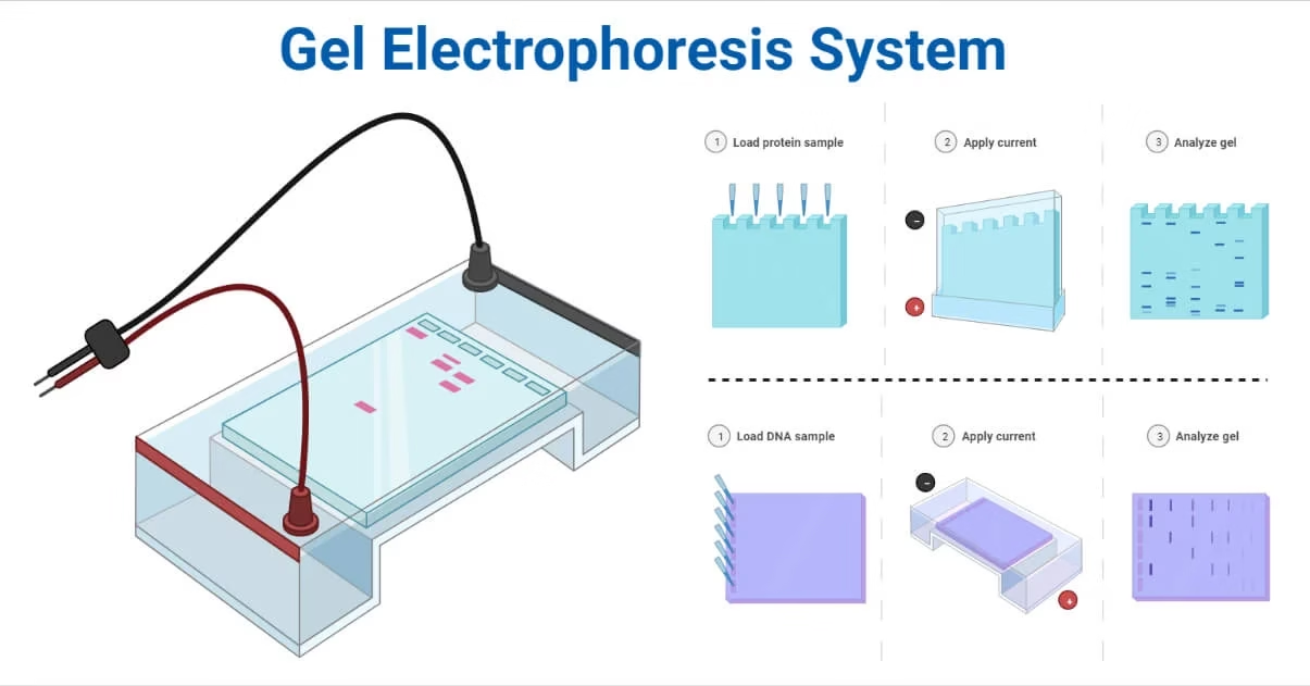
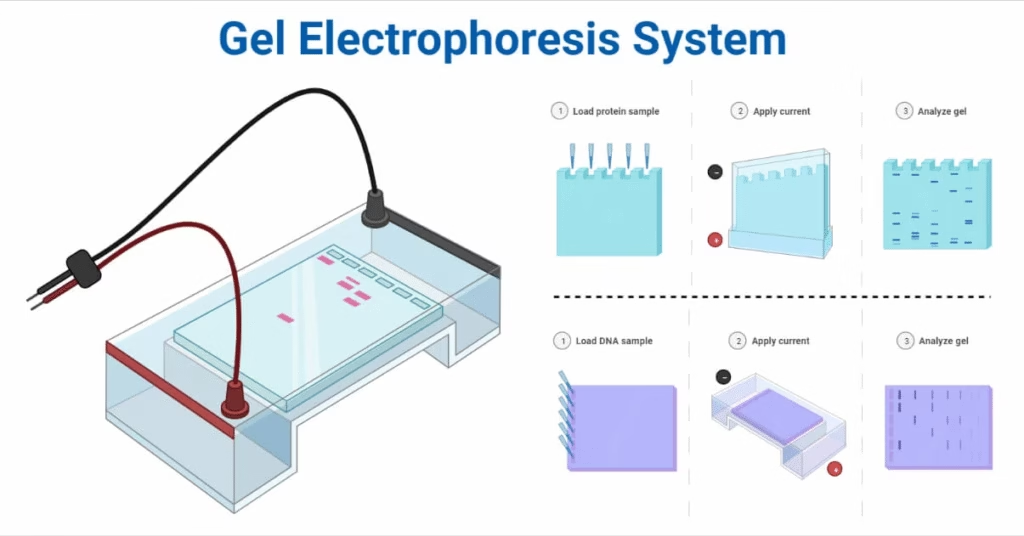
Operating Procedure of Gel Electrophoresis
- Gel Preparation – Dissolve agarose or acrylamide in buffer, heat, and pour into gel caster with comb.
- Gel Casting – Let gel solidify with wells created by comb.
- Sample Preparation – Mix DNA/protein with loading dye for visibility and density.
- Sample Loading – Pipette samples into wells.
- Running the Gel – Connect gel box to power supply. DNA (negative) moves towards anode.
- Stopping & Staining – Turn off current when bands are separated. Stain gel with EtBr, SYBR Green, or protein stains.
- Visualization – Use UV transilluminator (for DNA/RNA) or imaging systems. Compare bands with DNA ladder/marker.
Applications of Gel Electrophoresis
- Forensics – DNA fingerprinting for crime scene & paternity testing.
- Molecular Biology – DNA/RNA purification, cloning verification.
- Medicine – Identifying genetic mutations, protein disorders.
- Microbiology – Typing pathogens, studying microbial diversity.
- Evolutionary Biology – DNA profiling and taxonomy.
- Proteomics – Protein separation and identification.
- Vaccine Development – Purifying antigens and monitoring production.
- Blotting Techniques – Western blot (proteins), Southern blot (DNA), Northern blot (RNA).
Advantages of Gel Electrophoresis
- High resolution and accuracy.
- Can separate wide range of biomolecules.
- Relatively low cost.
- Easy to learn and perform.
- Compatible with multiple stains and detection methods.
- Provides both qualitative and semi-quantitative results.
Limitations of Gel Electrophoresis
- Requires large starting sample (especially proteins).
- Not suitable for small molecules like ions and hormones.
- Limited throughput (slower compared to PCR, sequencing).
- Cannot provide precise protein mass (requires MS).
- Sensitive molecules (heat-labile DNA/proteins) may degrade.
- Hazardous chemicals (EtBr, acrylamide) are used.
Precautions in Gel Electrophoresis
- Always wear gloves, goggles, lab coat.
- Handle Ethidium bromide and acrylamide carefully (toxic/mutagenic).
- Do not overload wells; use micropipette carefully.
- Avoid spills and short circuits in power supply.
- Use distilled water to prepare buffers.
- Store buffers cold to prevent bacterial growth.
Examples of Gel Electrophoresis Systems
- DNA Electrophoresis System GEP-TH-1000TBT (Bioevopeak)
- Built-in high-current power supply.
- Rapid transfer efficiency.
- Electrofocusing System BT105 (G Biosciences)
- No taping needed.
- Lightweight, easy to use.
- Mini DNA Electrophoresis EPS-2014 (Inovialab)
- Compact design for DNA/RNA.
- Safety lid with auto cut-off.
- SymphonyIEF Isoelectric Focusing (Hercuvan)
- Handles small to high-throughput IEF experiments.
FAQs on Gel Electrophoresis
Q1. What is gel electrophoresis?
Ans: Gel electrophoresis is a laboratory method used to separate DNA, RNA, or proteins based on their size, charge, and shape by applying an electric current through a gel matrix.
Q2. Who invented electrophoresis?
Ans: Electrophoresis was developed by Arne Tiselius in the 1930s. He was awarded the Nobel Prize in Chemistry in 1948 for his pioneering work.
Q3. What is the principle of gel electrophoresis?
Ans: Charged molecules migrate under an electric field:
- DNA/RNA (negatively charged) move towards the positive electrode (anode).
- Proteins may move towards positive or negative electrodes depending on their charge.
*Separation occurs because smaller molecules move faster through the gel pores.
Q4. Why is agarose gel used for DNA and RNA separation?
Ans: Agarose is a neutral, stable, and easy-to-handle polysaccharide with adjustable pore sizes, making it perfect for separating DNA/RNA fragments between 100 bp and 20 kb.
Q5. What is polyacrylamide gel electrophoresis (PAGE)?
Ans: PAGE is a high-resolution technique used for proteins and small nucleic acids. It can be run as native PAGE (proteins remain folded) or SDS-PAGE (proteins separated only by size).
Q6. What is SDS-PAGE used for?
Ans: SDS-PAGE uses the detergent Sodium Dodecyl Sulfate (SDS) to denature proteins and give them a uniform negative charge, allowing separation based purely on molecular weight.
Q7. What is pulsed-field gel electrophoresis (PFGE)?
Ans: PFGE is used to separate very large DNA fragments (up to millions of base pairs) by periodically changing the direction of the electric field. It is widely used in microbial typing and epidemiology.
Q8. What is a DNA ladder in gel electrophoresis?
Ans: A DNA ladder (or marker) is a set of DNA fragments of known sizes that helps in estimating the size of unknown DNA fragments during electrophoresis.
Q9. What are the main types of gel electrophoresis?
Ans: Major types include:
- Agarose gel electrophoresis (DNA/RNA)
- Polyacrylamide gel electrophoresis (PAGE)
- SDS-PAGE (proteins by size)
- Isoelectric focusing (proteins by pI)
- 2D gel electrophoresis (proteins by charge and size)
- PFGE (large DNA fragments)
- Immunoelectrophoresis (antigen-antibody studies)
Q10. Can RNA be separated by gel electrophoresis?
Ans: Yes, RNA can be separated by agarose gel electrophoresis, but special precautions (RNase-free conditions) are needed because RNA is easily degraded.
Q11. What is the difference between agarose gel and PAGE?
Ans:
- Agarose gel: Lower resolution, used for large DNA/RNA fragments.
- PAGE: High resolution, used for proteins and small nucleic acids.
Q12. Why do we add loading dye to DNA samples before electrophoresis?
Ans: Loading dye adds color (for visibility) and glycerol/sucrose (to make samples sink into wells). It also contains tracking dyes to monitor migration.
Q13. What is the purpose of using a buffer in electrophoresis?
Ans: Buffers maintain pH, ionic strength, and conductivity, ensuring proper migration of molecules. Common buffers are TAE, TBE, and Tris-glycine.
Q14. How are proteins visualized in gel electrophoresis?
Ans: Common stains include:
- Coomassie Brilliant Blue (general protein stain)
- Silver stain (very sensitive)
- Fluorescent dyes (for quantitative detection)
Q15. What is the difference between native and denaturing PAGE?
Ans:
- Native PAGE: Proteins remain in natural structure; separation based on size + charge.
- Denaturing PAGE (SDS-PAGE): Proteins are unfolded; separation only by size.
Q16. What are the applications of gel electrophoresis?
Ans: Applications include:
- DNA fingerprinting (forensics, paternity tests)
- Gene cloning verification
- Identifying mutations
- Protein purification and analysis
- Microbial typing (epidemiology)
- RNA analysis in gene expression studies
- Blotting techniques (Southern, Northern, Western blots)
Q17. What are the advantages of gel electrophoresis?
Ans:
- High accuracy and resolution
- Low cost and easy setup
- Can analyze multiple samples
- Suitable for DNA, RNA, and proteins
- Provides both qualitative and semi-quantitative data
Q18. What are the limitations of gel electrophoresis?
Ans:
- Requires relatively large samples
- Cannot analyze very small molecules (ions, hormones)
- Hazardous chemicals (EtBr, acrylamide) used
- Limited throughput compared to sequencing
- Some biomolecules may degrade during process
Q19. Why is ethidium bromide used in DNA electrophoresis?
Ans: Ethidium bromide (EtBr) intercalates between DNA bases and fluoresces under UV light, making DNA bands visible. However, it is toxic and mutagenic, so safer alternatives (SYBR Green, GelRed) are now preferred.
Q20. Can proteins be separated without SDS?
Ans: Yes, using native PAGE or isoelectric focusing. However, SDS-PAGE is preferred when studying protein molecular weights.
Q21. Why do small DNA fragments move faster in agarose gel?
Ans: Smaller fragments can pass through the pores of the gel more easily, while larger fragments are slowed down by resistance.
Q22. What precautions should be taken during gel electrophoresis?
Ans:
- Wear gloves and goggles.
- Handle EtBr and acrylamide carefully (toxic).
- Avoid air bubbles while casting gels.
- Do not overload wells.
- Ensure correct electrode connection (DNA runs towards positive electrode).
Q23. What is 2D gel electrophoresis used for?
Ans: 2D gel electrophoresis separates proteins in two dimensions – first by charge (isoelectric focusing) and then by size (SDS-PAGE). It is a major tool in proteomics research.
Conclusion
The Gel Electrophoresis System is one of the most powerful and widely used laboratory methods for separating, identifying, and analyzing biomolecules. From DNA fingerprinting in forensic science to protein analysis in medicine and biotechnology, it remains an indispensable tool in research and diagnostics.
Although it has some limitations (sample size, hazardous chemicals, low throughput), its simplicity, accuracy, and reliability make it a cornerstone technique in modern biology.
In short: Gel electrophoresis is the scientist’s microscope for DNA, RNA, and proteins.
References
- Viswanathan, S., Unlü, M., & Minden, J. S. (2006). Two-dimensional difference gel electrophoresis. Nature protocols, 1(3), 1351–1358. https://doi.org/10.1038/nprot.2006.234
- Büyükköroğlu, G., Dora, D. D., Özdemir, F., & Hızel, C. (2018). Techniques for Protein Analysis. Omics Technologies and Bio-Engineering, 317–351. doi:10.1016/b978-0-12-804659-3.00015-4
- https://javalab.org/en/dna-electrophoresis/
- https://conductscience.com/introduction-to-electrophoresis/
- https://study.com/learn/lesson/agarose-gel-electrophoresis-steps-purpose.html
- https://microbenotes.com/gel-electrophoresis-system-apparatus-parts-types-examples/
- https://www.vedantu.com/biology/sds-page
Other related topics you might be interested in:
Agarose Gel Electrophoresis – Principle, Procedure, Applications, Advantages & Limitations