The system that controls and coordinates all the activities of various body parts in response to external and internal stimuli by the conduction of nerve impulses is called nervous system.
Neuron or nerve cell
- It is the structural and functional unit of nervous system.
- A nerve cell with all its processes is called a neuron.
- It controls and integrates the different bodily functions and maintains the consistency of internal environment.
- Nerve cell respond to different stimuli.
- They have the property of irritability and conductivity.
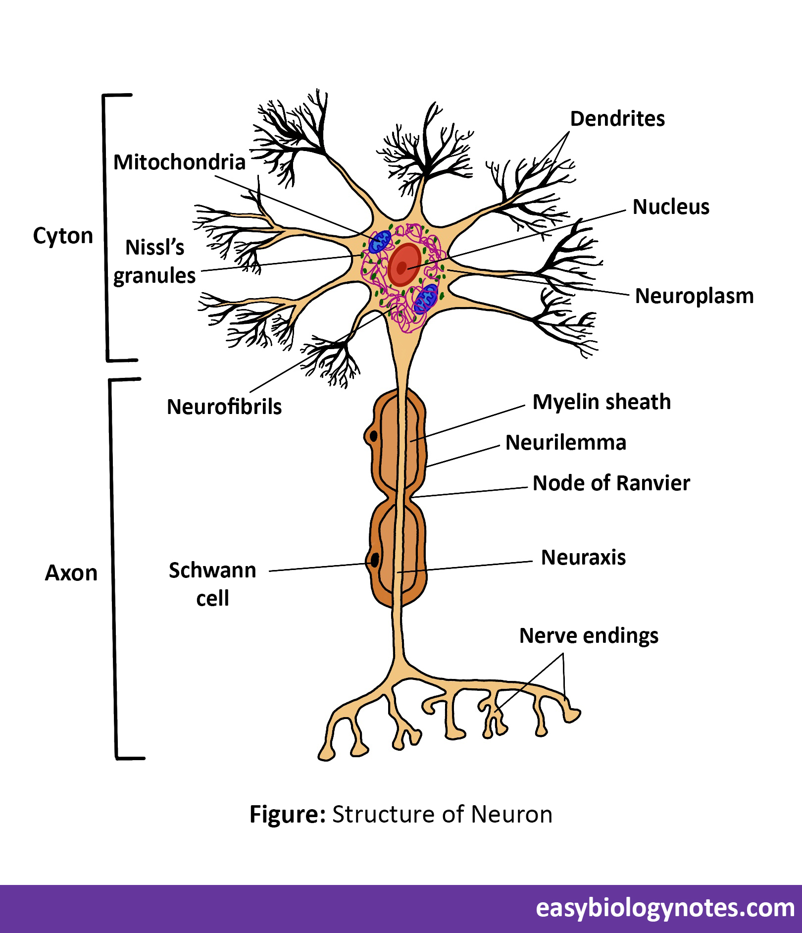
Structure of neuron
It consists of three main parts -:
- Cell body (Cyton or perikaryon)
- It contains a well defined nucleus and its cytoplasm is called neuroplasm.
- It also contains neurofibrils which help in the transmission of impulses.
- Centrosome are absent thus the neurons do not have the ability to divide.
- Neuroplasm also contain Nissl’s granules which are small fragments of rough endoplasmic reticulum.
- Also contains numerous mitochondria.
- Axon
- Also known as nerve fibre.
- Length varies from few millimeter to one metre.
- It is surrounded by a white insulating sheath known as neurilemma.
- Axon is a long slender projection of a nerve cell that conducts electrical impulses away from the cell body.
Types of nerves fibre
-
- An axon of a neuron covered by one or two sheaths is called nerve fibre.
- Depending upon the sheath’s covering of neuraxis (axis of axon) the nerve fibres are of two types:
- Myelinated nerve fibres (Medullated nerve fibres)
- In these nerve fibres the axon of neuron is surrounded by two sheaths – inner sheath known as myelin sheath while the outermost known as neurilemma.
- Myelinated nerve fibres show gaps known as nodes of Ranvier where myelin sheath is not continuous showing constrictions.
- Functions of Myelin Sheath
- To increase the speed of conduction of nerve impulse.
- To insulate the axon, i.e. ,prevents mixing of impulses in the adjacent fibres.
- Non-myelinated nerve fibre (Non-medullated nerve fibre)
- In these nerve fibre myelin sheath is absent and the axon is covered by only one sheath, neurilemma.
- Dendron or dendrites
- These are highly branched small projections arising from cyton.
- They conduct nerve impulses from synapses toward the cell body.
- Myelinated nerve fibres (Medullated nerve fibres)
Nerves
- Bundle of nerve fibres (axons) of separate neurons is covered by a tubular sheath (nerve sheath) known as nerve.
- There are three kinds of nerves -:
- Sensory nerves (Afferent nerves)
- Contains only sensory nerve fibres which bring impulses from receptors to the brain or spinal cord (CNS or Central Nervous System).
- Example – Optic nerve arising from the eye and ending in the brain.
- Motor nerves (Efferent nerves)
- Contains only motor nerve fibres which carry impulses from Central Nervous System to the effector organ.
- Example – A nerve arising from the brain connected to the muscles of eyeball in order to rotate the eye.
- Mixed / Association / Connector nerves
- It contains both sensory and motor nerve fibres.
- Sensory nerves (Afferent nerves)

Synapse
- Nerve signals are transmitted from one neuron to the next through the interneural junctions called synapse. Synapse is a point of contact where two neurons are functionally connected.
- The terminal branches of the axon of one neuron come in contact with the dendrites or the cell body or the axon part of another neuron.
- The gap between the dendrite and the axon terminal of a synapse is called synaptic cleft.
Neurotransmitter
- These are the chemical messengers produced by the nervous system that transmits signal from a neuron to another cell across a synapse.
- For example – Acetylcholine which get broken down by an enzyme to make the synapse ready for the next transmission.

NERVOUS SYSTEM
The nervous system is a network of nerves and other tissues that controls and coordinates the body’s functions. It refers to the brain, nerves, spinal cord, ganglia and other receptor organs that receive and interpret stimuli.
Functions of nervous system
- It controls all the functions of various body parts.
- It coordinates and integrates activities of all the body parts.
- It controls all the voluntary muscular activities.
- It regulates involuntary activities.
- It enables us to remember, to think and to reason out.
- It controls all the reflex actions.
*Conduction of nerve impulse is a wave of depolarization followed by repolarization.

A. Central Nervous System (CNS)
All the parts of the nervous system which are present along the median longitudinal axis of the body constitute the CNS. It consists of two parts –:
1. Brain
- Position – Brain is a very delicate organ well protected inside the cranial cavity or brain box of the skull (cranium).
- Average weight is of about 1350g.
- Human brain is made of about 1000 billion neurons.
- Protections and coverings :
- Dura mater – Outermost thick, non-vascular, tough fibrous membrane.
- Arachnid – Thin delicate middle layer, non-vascular giving a web-like cushion.
- Pia mater – Innermost, highly vascular, thin and tough membrane, richly supplied with blood. It nourishes the brain.
Structure of Brain


Human brain is whitish, bilaterally symmetrical structure which is divisible into three regions -:
-
-
-
-
- Forebrain
-
-
-
It is differentiated into three parts :
a. Olfactory lobes – Olfactory lobes are a pair of poorly developed, club-shaped, widely separated bodies.
- They are visible from the ventral surface only.
- These are concerned with the sense of smell.
b. Cerebrum – It is the largest part which forms 4/5th of the weight of brain.
- Cerebrum is divided into right and left lobes called cerebral hemispheres.
- Their surface is highly folded or convoluted with ridges (Gyrus : gyri) and grooves (Sulcus : sulci) to increase the surface area to accommodate more neurons and hence greater intelligence.
- Each cerebral hemisphere is hollow from inside and the walls have two regions – Outer cortex and inner medulla.
- The outer portion of cerebrum (cortex) contains grey colored cell bodies of the neurons and hence called grey matter which is folded to form convolutions.
- The inner portion of cerebrum (medulla) consists of axons of neurons which is white in color and hence called white matter.
- *Cerebral fissure – It is a very deep groove which separates two cerebral hemispheres from each other.
- *Corpus callosum – It is a sheet of nerve fibres connecting the two cerebral hemispheres. Its function is to transfer information from one hemisphere to other.
Functions of Cerebrum
- It is the seat of memory, intelligence, will power, emotions, experiences, reasoning, learning, invent and plan.
- It initiates and controls all voluntary actions by controlling the movement of skeletal muscles.
- It perceives the sensory impulses such as pain, touch, smell, hearing and site.
- Subconscious or unconscious part is also located in the cerebrum.
c. Diencephalon – It is a small rhomboidal lobe.
- It is a small rhomboidal lobe.
- It is completely covered superiorly by large cerebrum. It lies between cerebrum and the mid brain.
It has two parts -:
- Thalamus – Acts as a relay centre for pain and pressure impulses to the cerebrum.
- Hypothalamus – Controls the body temperature and pituitary and also the blood pressure.
2. Midbrain
- It is a small tubular part which connects the forebrain with the cerebellum and pons of hindbrain.
Functions of midbrain
- It conveys impulses from hindbrain to forebrain.
- It controls the sight and auditory impulses.
3. Hindbrain – It is the posterior smallest part of the brain which is differentiated into three parts -:
a. Cerebellum
- It is the largest part of the hindbrain located just at the base of cerebrum and above the medulla oblongata.
- It consists of two large lateral lobes called cerebellar.
- It has no convolutions but has numerous furrows giving it a laminated appearance.
- It has an inner core and white matter surrounded by grey matter.
Functions of cerebellum
- It helps to maintain the balance or equilibrium during movements.
- It controls and coordinates muscular activities.
b. Medulla oblongata
- It is the lowest portion of the brain located at the base of the skull.
- It is about 2.5 cm long, roughly triangular extending from pons to spinal cord.
c. Pons varolii
- It is located in the centre of the brain below the cerebellum.
Functions -:
- It carries impulses from one hemisphere of the cerebellum to the other hemisphere of the cerebellum and coordinates muscular movements on both the sides of the body.
2. Spinal cord
- It is a long unsegmented cord-like structure extending from medulla oblongata of the brain through the neural canal of the vertebral column to the lumbar region.
- It is about 45 cm in length and 35 gm in weight.
- It is also covered by three membranes known as meninges.
- The arrangement of white and grey matter is reversed from that in the brain, i.e. , white matter is external and grey matter is internal in position.
- The spinal cord is hollow from inside containing a cavity called central canal which runs through the entire length of spinal cord.
- Central canal is filled with cerebrospinal fluid (CSF) which acts as a shockproof cushion and forms a medium for the exchange of food materials, waste products and respiratory gases with neurons.

Functions of spinal cord
- It controls reflexes below the neck.
- It functions as reflex centre which controls all the spinal reflexes.
- Conducts sensory impulses from skin to the brain and motor responses from brain to the skin.
B. Peripheral Nervous System (PNS)
All the whitish, thread-like nerves which connect the various body parts with the CNS collectively constitute the PNS. It is divided into two parts –:
1. Somatic Nervous System
It consists of cranial and spinal nerves.
-
- Cranial nerves
- Those nerves which arise from the brain are called cranial nerves.
- There are 12 pairs of cranial nerves.
- The cranial nerves may be sensory – Like olfactory nerves (nose), optic nerves (eye) , auditory nerves (ear) ; or motor nerves – Oculomotor nerve (going to the eye muscles); and mixed in nature – facial nerves.
- Spinal nerves
- Nerves arising from the spinal cord are called spinal nerves.
- There are 31 pairs of spinal nerves.
- Every spinal nerve is a mixed nerve having both sensory and motor fibres.
- At the junction of the two roots the sensory and the motor fibres separate out – the sensory fibres continue in the dorsal root and the motor fibres into the ventral root.
- Spinal nerves are classified into the following five groups :
- Cervical spinal nerves in the neck – 8 pairs
- Thoracic spinal nerves in thorax – 12 pairs
- Lumbar spinal nerves in abdomen – 5 pairs
- Sacral spinal nerves in hip region – 5 pairs
- Coccygeal spinal nerves in tail region – 1 pair
- Cranial nerves
2. Autonomic Nervous System (ANS)
- It includes a pair of chains of ganglia (aggregates of cell body of neurons) and nerves which control the involuntary actions of many internal organs (smooth muscles, cardiac muscles and glands) .
- That part of the nervous system which controls all the involuntary activities of various body parts is called ANS.
- It is divided into two parts :
1. Sympathetic Nervous System
- It prepares the body for violent action during emergency.
- It is stimulated by the hormone – Adrenaline .
- Nerves of sympathetic system arise from spinal cord between the neck and the wrist region.
2. Parasympathetic Nervous System
- It helps in reestablishing normal conditions after the violent act is over.
- It is stimulated by the hormone – Noradrenaline .
- Nerves of parasympathetic system are located at two places – one in the head and the neck and other in the sacral region.
Difference between Sympathetic and Parasympathetic Nervous System
| Sympathetic Nervous System | Parasympathetic Nervous System | |
| 1. | Dilates pupils of eyes. | Constricts pupils of eyes. |
| 2. | Increases heartbeat. | Decreases heartbeat. |
| 3. | Increases breathing by dilating bronchi and bronchioles. | Decreases breathing by constricting bronchi and bronchioles. |
| 4. | Stimulates tear secretion. | Inhibits tear secretion. |
| 5. | Increases blood pressure. | Decreases blood pressure. |
| 6. | Dilates nostrils. | Constricts nostrils. |
| 7. | Relaxes urinary bladder. | Contracts urinary bladder. |
| 8. | Decreases secretion of saliva. | Increases secretion of saliva. |
| 9. | Decreases secretion of digestive glands. | Increases secretion of digestive glands. |
| 10. | Decreases peristalsis in gut. | Increases peristalsis in gut. |
Reflexes
- Reflex action is a spontaneous, immediate, involuntary and automatic response to any stimulus without the involvement of the brain.
- Reflex actions are performed unconsciously.
- Examples :
- Flushing of tears
- Instantaneous withdrawal of hand when it touches a hot iron.
- Shivering or sweating.
- Dilation of the eye pupil.
Types of reflexes
There are two types of reflexes – Unconditional reflexes and Conditional reflexes
* Unconditional reflexes are innate, natural , inborn or simple reflexes.
* Conditioned reflexes are acquired reflexes.
| Unconditioned reflexes | Conditioned reflexes | |
| 1. | They are specific ,i.e. , they are the characteristics of all members of a given species. | They are individual and may be present in some members of a species and absent in others. |
| 2. | Unconditional reflexes are relatively stable. | They are unstable and may be developed, reinforced or extinguished under definite conditions , as properly reflected in their very name. |
| 3. | They are involved in response to adequate stimuli applied to definite respective fields. | They can be developed under the action of the most varied stimuli applied to different respective fields. |
Mechanism of unconditioned reflexes
Reflex arc – The path covered by the impulses from receptor to the effector – through sensory or afferent nerve fibres, a part of CNS, and efferent or motor nerve fibre – is known as reflex arc.
Conditioned reflexes
- A conditioned reflex is an involuntary, spontaneous, automatic response brought about due to a previously learnt experience.
- The idea of conditioned reflex action was given by Pavlov through an experiment on a dog.
- He demonstrated that a particular response could be evoked to a particular stimulus that does now occur in nature.
- Pavlov called sound of the bell as conditioned stimulus and salivation in response to bell as conditioned response, whereas the food itself is an unconditioned stimulus and salivation in response to food is an unconditioned response.
Pavlov’s experiment
Food 🡪 Dog salivates
Ringing of bell 🡪 Dog does not salivate
Ringing of bell along with the presentation of food 🡪 Dog salivates
Ringing the bell (not followed by food) 🡪 Dog salivates
Significance of reflexes
- They are spontaneous response to harmful stimulus, thus protecting us from the harm.
- They relief the burden of brain by automatically responding to stimuli.
- Most of the daily habits are conditioned reflexes.
Some Key terms
Ependyma – Epithelial linings of the cavities found in parts of brain and spinal cord.
Impulse – A wave of change in the electric potential on the outer and inner side of nerve fibre.
Irritability – Response to external stimuli.
Response – Change or reaction of the body due to stimulus.
Stimulus – Any agent or change in the environment that can cause change or reaction in the body is called stimulus.
Myelitis – It is a medical term for inflammation of spinal cord.